
Gallstones
11 Sept 2026 · 18 min read
On this page43 sections
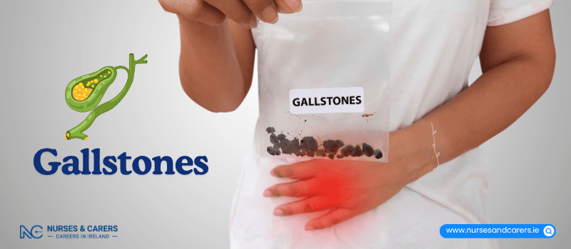
Gallstones are solid, crystal-like deposits that form inside the gallbladder. They most commonly develop from hardened cholesterol, a type of lipid found naturally in your bloodstream.
In the majority of cases, gallstones remain asymptomatic and require no medical intervention. However, they can lead to complications if they shift and block the flow of bile. This can result in biliary colic (intermittent abdominal pain) or cholecystitis (acute inflammation of the gallbladder). When gallstones cause noticeable symptoms or clinical complications, the condition is medically classified as gallstone disease or cholelithiasis.
Anatomy and Function of the Gallbladder
The gallbladder is a small, pouch-like organ located directly beneath the liver. It does not produce bile; rather, it serves as a storage reservoir for it. Bile is synthesized by the liver and travels through a network of channels called bile ducts into the gallbladder. Its primary function is to break down and help the body digest dietary fats.
If the gallbladder needs to be surgically removed, you can continue to live a perfectly normal, healthy life. The liver will still produce an adequate amount of bile to digest food, draining it directly into the small intestine instead.
Causes and Risk Factors
Gallstones typically form due to a chemical imbalance in the composition of the bile. When the concentration of cholesterol within the bile becomes excessively high, the surplus crystallizes and hardens into stones.
Gallstones are highly prevalent, affecting approximately 1 in 10 adults, though most individuals remain completely unaware of them. Key factors that elevate your risk of developing gallstones include:
Carrying excess body weight or having obesity
Being biologically female, with an increased risk for those who have carried pregnancies
Being 40 years of age or older, as the risk steadily rises across the lifespan
Treatment Options
Medical treatment is generally reserved only for individuals experiencing active symptoms or related complications, such as jaundice (yellowing of the skin and eyes) or acute pancreatitis (inflammation of the pancreas).
When treatment is necessary, physicians typically recommend keyhole surgery to remove the organ entirely. This routine surgical procedure is known as a laparoscopic cholecystectomy. The vast majority of gallstone disease cases are successfully resolved through this safe and common operation.
Overview of Symptoms
In the majority of cases, gallstones remain completely asymptomatic. However, symptoms can manifest over time if a gallstone shifts and obstructs one of the bile ducts. This blockage typically triggers sudden, severe abdominal pain known as biliary colic. If the obstruction becomes prolonged or spreads to other areas of the digestive system, additional clinical signs can develop.
Abdominal Pain (Biliary Colic)
The defining symptom of gallstones is sudden, intense abdominal pain. This pain is often description-based as cramping or colicky, meaning its intensity may fluctuate.
Duration: An episode typically lasts between 1 and 5 hours, though it can occasionally resolve within a few minutes.
Location: The pain is localized in the upper abdomen, specifically just beneath the ribs on the right side. From there, it may radiate through to the right shoulder blade.
Triggers and Timing: While it can occur at any time—including waking you up in the middle of the night—it is frequently triggered by eating fatty foods.
Relief: Unlike standard digestive discomfort, this pain is not relieved by using the restroom, passing gas, or vomiting.
These episodes are usually infrequent, with weeks or months passing between occurrences. During an attack, you may also experience profuse sweating, nausea, or vomiting. When gallstones cause these isolated episodes of pain without further illness, the condition is classified as uncomplicated gallstone disease.
Signs of Complicated Gallstone Disease
A small percentage of individuals may develop more severe issues if a gallstone completely blocks the bile duct for an extended period or migrates into neighboring organs, such as the pancreas or small intestine. This progression is known as complicated gallstone disease and can present with the following symptoms:
A persistent high temperature (38°C / 100.4°F or above)
Constant, unremitting abdominal pain
A rapid heart rate (tachycardia)
Yellowing of the skin and the whites of the eyes (jaundice)
Persistently itchy skin
Diarrhea
Chills or episodes of shivering
Unexplained confusion or disorientation
A sudden loss of appetite
When to Seek Medical Attention
Non-Urgent Care
Schedule an appointment to see your General Practitioner (GP) if you suspect you are experiencing intermittent abdominal pain related to gallstones (biliary colic).
Urgent Medical Care
Contact your GP urgently or seek immediate emergency medical care if you develop any of the following warning signs:
Jaundice: Visible yellowing of your skin or eyes.
Severe, Prolonged Pain: Abdominal pain that lasts for more than 8 hours, or pain so intense that you cannot find relief in any sitting or lying position.
Infection Signs: A high fever accompanied by chills and shivering.
Etiology and Chemical Imbalances
Gallstones develop due to a chemical imbalance in the composition of bile—the digestive fluid produced by the liver and stored in the gallbladder. While the precise triggers for this imbalance remain unclear, stones form when certain substances precipitate and crystallize.
There are two primary types of gallstones based on their composition:
Cholesterol Gallstones: Account for approximately 80% (4 out of 5) of all cases. They form when the liver excretes more cholesterol than the bile can dissolve.
Pigment (Bilirubin) Gallstones: Account for approximately 20% (1 out of 5) of cases. Bilirubin is a natural waste product created when the body breaks down old red blood cells. Certain conditions can cause the liver to produce too much bilirubin, leading to stone formation.
This crystallization process is gradual and can take several years. Gallstones vary significantly in size, ranging from microscopic grains of sand to the size of a pebble. An individual may develop a solitary stone, but it is far more common to have multiple stones present simultaneously.
Risk Factors
While anyone can develop gallstones, certain demographic, genetic, and medical factors significantly increase your susceptibility.
Demographic and Biological Factors
Biological Sex: Gallstones are notably more common in females. Risk increases further with pregnancy, the use of the combined oral contraceptive pill, or high-dose estrogen hormone therapy.
Age: The risk steadily escalates with age, becoming much more prevalent in individuals aged 40 and older.
Genetics: Having a close family member with a history of gallstones increases your personal risk.
Weight and Metabolic Factors
Body Weight: Carrying excess weight or living with obesity elevates the concentration of cholesterol in your bile.
Rapid Weight Loss: Losing weight quickly—whether through intensive dieting or bariatric (weight-loss) surgery—can alter bile chemistry and cause the gallbladder to empty less effectively, triggering stone formation.
Medical Conditions and Medications
Gastrointestinal Disorders: Living with Crohn's disease alters how your body absorbs bile salts, increasing risk.
Medications: Using certain medications, such as the antibiotic ceftriaxone, can promote stone formation.
Impaired Bile Flow: Any underlying health condition that restricts or slows the natural flow of bile out of the liver or gallbladder.
Specific clinical conditions that impair bile flow include:
Cirrhosis: Advanced scarring of the liver tissue.
Primary Sclerosing Cholangitis (PSC): Chronic inflammation and scarring of the bile ducts.
Intrahepatic Cholestasis of Pregnancy (Obstetric Cholestasis): A liver condition that can develop during pregnancy, slowing the flow of bile.
Initial Assessment by Your GP
Gallstones are frequently discovered incidentally during diagnostic imaging for an unrelated medical issue, particularly when they are asymptomatic. However, if you develop symptoms, you should schedule an appointment with your General Practitioner (GP) for a clinical evaluation.
During your consultation, your GP will review your medical history and discuss your symptoms. They will perform a targeted physical examination to check for gallbladder inflammation, which may include Murphy's sign:
The Examination: The doctor will place their hand on the upper-right quadrant of your abdomen and apply gentle pressure.
The Response: If you experience sharp pain or find it difficult to take a full breath in during this pressure, it typically indicates an inflamed gallbladder. This finding may require urgent medical care.
Your GP may also request blood tests to screen for biochemical markers of an active infection and to evaluate your liver function. Gallstones that migrate into the bile ducts can obstruct the flow of bile, directly impacting liver enzyme levels.
Advanced Diagnostic Imaging
If your initial physical assessment or blood panels suggest the presence of gallstones, your GP will refer you for further diagnostic testing. These non-invasive procedures are typically performed in a hospital outpatient department, though individuals with severe symptoms may be admitted to the hospital for immediate evaluation.
1. Abdominal Ultrasound Scan
An ultrasound scan is the standard diagnostic tool used to confirm the presence of gallstones. It is a completely painless procedure that takes approximately 10 to 15 minutes.
The Process: A sonographer applies a conductive gel to your abdomen and moves a handheld device (transducer) across the skin. This device utilizes high-frequency sound waves to generate live images of your internal organs onto a monitor.
Limitations: While an ultrasound is highly effective at identifying stones within the gallbladder itself, it cannot always definitively see smaller stones that have migrated into the bile ducts.
2. Magnetic Resonance Cholangiopancreatography (MRCP)
If your clinical team suspects that gallstones have moved into your bile ducts, they will recommend a specialized type of MRI scan known as an MRCP.
The Process: You will lie inside a cylindrical, tube-shaped MRI scanner. The machine uses a strong magnetic field and radio waves to generate highly detailed, high-resolution cross-sectional images of your bile ducts, gallbladder, and pancreatic duct.
3. Computed Tomography (CT) Scan
A CT scan is primarily utilized to evaluate potential complications of gallstone disease, such as a perforated gallbladder, abscess, or acute pancreatitis.
The Process: The scanner resembles a large, open-ended doughnut that captures a rapid series of X-ray images from multiple angles.
Clinical Use: Because CT scanners can quickly capture detailed structural data, they are frequently utilized in emergency departments to rapidly diagnose the cause of severe, acute abdominal pain.
Clinical Management Strategies
The treatment plan for gallstones depends on whether you have symptoms and how severely they impact your daily life. If your gallstones are asymptomatic, medical intervention is generally not required. However, you should monitor your health and contact your General Practitioner (GP) if symptoms develop.
Proactive Treatment for At-Risk Individuals
In specific cases, doctors recommend treating asymptomatic gallstones if an underlying health condition increases your risk of severe complications. These conditions include:
Cirrhosis: Advanced scarring of the liver.
Portal Hypertension: High blood pressure within the liver's blood vessels, frequently caused by alcohol-related liver disease.
Diabetes: A metabolic condition that elevates the risk of rapid infection.
Gallbladder Calcification: High levels of calcium deposits in the gallbladder wall (often visible on a scan), which can increase the long-term risk of gallbladder cancer
Non-Surgical Management and Diet
For mild, infrequent episodes of abdominal pain (biliary colic), surgical intervention may not be immediately necessary.
Pain Management: Your GP can prescribe or recommend appropriate painkillers to manage acute discomfort.
Dietary Adjustments: Eating a balanced, low-fat diet can help reduce the frequency of gallbladder contractions and control pain. Avoiding known triggers—such as highly fatty, greasy, or heavily spiced foods—can also minimize unpleasant secondary symptoms like bloating and diarrhea.
While dietary changes improve overall health and manage symptoms, they do not cure gallstones or dissolve existing ones.
Surgical Interventions
Because the gallbladder is not an essential organ, removing it is a safe and permanent solution to gallstone disease. Your liver will continue to produce bile, allowing you to lead a normal, healthy life.
1. Keyhole Surgery (Laparoscopic Cholecystectomy)
This is the standard and most common surgical method for gallbladder removal.
Pre-Surgery: The procedure is performed under general anesthesia, ensuring you are completely asleep and pain-free. The operation typically takes 60 to 90 minutes.
The Procedure: The surgeon makes 3 or 4 minor incisions in your abdomen, including one slightly larger incision (2cm to 3cm) near your belly button. Your abdomen is inflated with harmless carbon dioxide gas to optimize visibility. Using a laparoscope (a thin tube with a camera and light source) and specialized miniature instruments, the surgeon removes the gallbladder. They may also take an intraoperative X-ray or ultrasound to check for and clear any hidden stones in the bile ducts.
Post-Surgery: Most individuals return home the same day. Full recovery generally takes about 10 days. Note: You may experience temporary shoulder discomfort post-surgery due to the carbon dioxide gas used during the procedure.
2. Open Surgery (Open Cholecystectomy)
If keyhole surgery is technically challenging or if an unexpected complication occurs, an open operation is performed. Your surgical team may advise against keyhole surgery from the outset if you:
Are in your third trimester of pregnancy.
Live with severe obesity.
Have an unusual anatomy of the gallbladder or bile ducts.
The Procedure: Performed under general anesthesia, the surgeon creates a larger 10cm to 15cm incision just below your ribs on the right side to directly access and remove the organ.
Post-Surgery: Open surgery is highly effective but requires a hospital stay of up to 5 days and a longer recovery time of approximately 6 weeks. It also leaves a more prominent permanent scar.
Alternative Treatments
1. Endoscopic Retrograde Cholangio-Pancreatography (ERCP)
An ERCP is a specialized procedure used exclusively to clear gallstones from the bile ducts. It does not remove the gallbladder itself; any stones remaining inside the gallbladder will persist.
The Procedure: You are given a sedative to ensure comfort while remaining awake. The surgeon passes a flexible endoscope down your throat and into the small intestine where the bile duct opens. They then widen the duct opening using a small incision or heated wire to remove the stones or allow them to pass safely into your digestive tract. A temporary plastic or metal tube called a stent may be inserted to keep the duct open.
Post-Surgery: The procedure takes 15 to 60 minutes. An overnight hospital stay is standard to monitor your recovery.
2. Oral Medication
In very rare instances, specific oral medications can be prescribed to dissolve small, non-calcified cholesterol gallstones. This treatment is rarely utilized because:
It has a low success rate.
It requires continuous, daily medication for up to 2 years.
Gallstones frequently return once the medication is stopped.
Overview of Complications
While most gallstones do not cause issues, a small percentage of individuals may experience severe complications. These generally happen when a stone causes a prolonged blockage in a duct or migrates into another area of the digestive tract.
1. Acute Cholecystitis (Gallbladder Inflammation)
If a gallstone permanently blocks the cystic duct, bile builds up inside the gallbladder. This stagnation can lead to infection and severe inflammation of the organ walls.
Key Risk: If left untreated, an inflamed gallbladder can lead to serious localized infections or tissue death (necrosis).
2. Jaundice
Jaundice occurs when a gallstone exits the gallbladder and obstructs the common bile duct, blocking the natural flow of bile into the intestines. This causes a yellow pigment called bilirubin to build up in the bloodstream.
Clinical Signs: Yellowing of the skin and the whites of the eyes, persistently itchy skin, dark brown urine, and pale or clay-colored stools.
Management: While some stones pass spontaneously, many require medical removal to prevent liver damage.
3. Acute Cholangitis (Bile Duct Infection)
When a gallstone creates a persistent blockage in the bile ducts, bacteria can rapidly multiply, causing a life-threatening bacterial infection known as acute cholangitis.
Symptoms: Severe upper abdominal pain radiating to the shoulder blade, a high fever (38°C/100.4°F or above), chills, jaundice, dark urine, pale stools, confusion, and general malaise.
Treatment: This is a medical emergency. Treatment requires intravenous antibiotics and immediate bile drainage, typically performed via an Endoscopic Retrograde Cholangio-Pancreatography (ERCP).
4. Acute Pancreatitis
This condition develops if a migrating gallstone blocks the pancreatic duct, preventing digestive enzymes from leaving the pancreas and causing the organ to become inflamed.
Symptoms: A sudden, severe, and constant dull pain in the upper abdomen that often radiates through to the back. The pain worsens after eating but may ease slightly when leaning forward or curling into a fetal position. Secondary signs include nausea, vomiting, diarrhea, loss of appetite, fever, and abdominal tenderness.
Treatment: There is no direct cure; hospital care focuses on supportive therapy until the inflammation subsides. This includes intravenous fluids, intensive pain relief, nutritional support, and supplemental oxygen. Most patients recover and can leave the hospital within 5 to 10 days.
5. Gallbladder Cancer
Gallbladder cancer is an exceptionally rare malignancy, affecting fewer than 1 in 10,000 people. However, approximately 80% of individuals diagnosed with this cancer have a history of gallstones.
Preventative Removal: Your doctor may recommend a preventative cholecystectomy if you have a family history of gallbladder cancer, a "porcelain gallbladder" (heavy calcium deposits in the wall), or gallbladder polyps larger than 1.5 cm.
Treatment: Managed using a combination of oncological surgery, chemotherapy, and radiotherapy.
6. Gallstone Ileus
Gallstone ileus is a rare, life-threatening form of mechanical bowel obstruction. It occurs when a large gallstone erodes through the gallbladder wall, creating an abnormal channel (fistula) directly into the small intestine, where it eventually becomes trapped and blocks the bowel.
Symptoms: Severe abdominal pain, vomiting, abdominal swelling (distension), constipation, and an inability to pass gas.
Key Risk: If left untreated, the pressure can cause the bowel wall to rupture (tear), leading to life-threatening internal infections (peritonitis).
Treatment: Requires immediate emergency surgery to remove the obstructing stone and clear the bowel.
Emergency Warning Signs
Seek urgent medical attention if you experience any signs of a bowel obstruction (inability to pass gas or stool accompanied by vomiting), a high fever with shaking chills, or a sudden yellowing of your skin and eyes.
Lifestyle and Dietary Modifications
While there is no definitive method to completely prevent the formation of gallstones, you can significantly reduce your risk by managing metabolic factors and making specific dietary choices. Because the majority of gallstones are composed of cholesterol, prevention strategies primarily focus on regulating cholesterol levels and maintaining a healthy metabolism.
1. Dietary Adjustments
Lowering your intake of foods high in saturated fats helps prevent the liver from excreting excessive amounts of cholesterol into your bile.
Foods to Limit or Avoid:
Fatty Meats: Sausages, bacon, and highly marbled or fatty cuts of meat.
Solid Fats: Butter, ghee, lard, and suet.
Full-Fat Dairy: Heavy cream, sour cream, and hard cheeses (such as cheddar or parmesan).
Processed Sweets: Commercial cakes, pastries, and biscuits.
Specific Plant Oils: Foods prepared with coconut oil or palm oil, which are high in saturated fats.
Adopting a balanced diet rich in soluble fiber (such as oats, beans, and lentils), whole grains, and healthy unsaturated fats (like olive oil) supports optimal gallbladder function and healthy cholesterol levels.
2. Weight Management and Physical Activity
Living with excess body weight or obesity elevates the concentration of cholesterol in your bile, directly increasing your susceptibility to stone formation.
Sustainable Weight Control Involves:
Nutrient-Dense Eating: Focusing on portion control and whole foods rather than restrictive dieting.
Active Lifestyle: Engaging in regular physical activity, aiming for a mix of cardiovascular exercise and strength training to support metabolic health.
The Risk of Rapid Weight Loss
It is critical to avoid crash diets, extremely low-calorie regimens, or rapid weight-loss schemes. When the body loses weight too quickly, the liver secretes extra cholesterol into the bile, and the gallbladder may fail to contract properly. This disruption in bile chemistry dramatically increases, rather than decreases, the risk of developing gallstones.
For safe prevention, aim for a gradual, steady weight-loss target of approximately 0.5 to 1 kilogram (1 to 2 pounds) per week.
Disclaimer
The information provided here is for educational and informational purposes only. It does not constitute medical advice, diagnosis, or treatment. Always consult a qualified healthcare professional or your GP for specific medical concerns, diagnoses, or health advice. Never disregard professional medical advice or delay seeking it because of information you have read here.