
Kidney Stones (Nephrolithiasis)
12 Sept 2026 · 15 min read
On this page3 sections
Kidney stones develop when mineral waste products in the bloodstream form solid crystals that accumulate and consolidate inside the kidneys. Over time, these crystals clump together to form hard, stone-like masses. Kidney stones are highly prevalent and can develop at any age, though they occur most frequently in adults between the ages of 30 and 60.
Sensation and Symptoms
The medical term for kidney stones is nephrolithiasis. When a stone moves or causes an obstruction, it can trigger an acute episode of agonizing, spasmodic pain known as renal colic.
Small stones: Often produce no clinical symptoms. They frequently drift through the urinary tract unnoticed and exit the body naturally during urination.
Large stones: Can become structurally lodged inside the urinary system. They typically cause a blockage in the ureter (the narrow tube connecting the kidney to the bladder) or, rarely, the urethra (the exit tube).
A urinary blockage triggers severe, sharp pain in the abdomen, lower back, flank, or groin. It can also cause secondary complications, such as a urinary tract infection (UTI).
Common Root Causes
Stones form when your urine contains a high concentration of crystalline substances—such as calcium, oxalate, and uric acid—and lacks the fluid volume necessary to dilute them. This crystallization is significantly more likely to happen if you:
Chronically fail to drink enough hydrating fluids throughout the day.
Take certain prescription or over-the-counter medications that alter urine chemistry.
Have an underlying metabolic or medical condition that causes your body to excrete unusually high levels of minerals in your urine.
Prevention Strategies
The most effective way to prevent kidney stones is to maintain consistent, optimal hydration. Drinking plenty of fresh water every day dilutes your urine, which physically stops waste minerals from binding together into crystals.
Statistical Risk Note: If you have developed a kidney stone once, you have a high statistical probability of developing another. Roughly 50% of individuals who experience a kidney stone will experience a recurrence within 5 years. Strict fluid management is essential to lower this risk.
Clinical Diagnostics and Treatment
Most kidney stones are small enough to be flushed out naturally. If a stone is small, your doctor will typically advise you to manage the symptoms at home using prescribed pain relievers, anti-nausea medications, and plenty of oral fluids.
However, hospital admission and advanced medical intervention are required for large or complicating stones. Hospital therapies include:
Extracorporeal Shock Wave Lithotripsy (ESWL): Utilizing targeted ultrasound shock waves to safely shatter large stones into tiny fragments from outside the body, allowing them to pass naturally.
Laser Lithotripsy: Routing a micro-scope into the urinary tract to blast the stones apart using a precise laser beam.
Percutaneous Nephrolithotomy (PCNL): Minimally invasive keyhole surgery performed to physically extract very large or complex stones directly from the kidney.
Function of the Kidneys
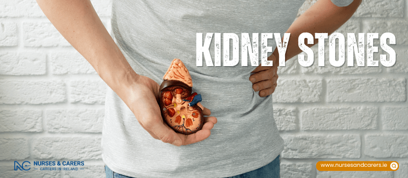
The kidneys are two bean-shaped organs, each measuring approximately 10cm (4 inches) in length, located just below the ribcage on either side of the spine. Their primary biological function is to continuously filter and remove waste products, excess fluids, and toxins from your bloodstream. The purified blood is returned to your circulatory system, while the filtered waste products are systematically excreted from the body as urine.
Symptoms of Kidney Stones
Small kidney stones often cause no symptoms at all. They can travel through the urinary tract completely unnoticed and pass out painlessly during urination. However, larger stones—or stones that shift and travel down the urinary system—can trigger severe symptoms.
Core Symptoms of a Kidney Stone
When a kidney stone moves or begins to obstruct the flow of urine, it can cause:
Severe, fluctuating pain: Agonizing pain located in your side (flank), lower abdomen, or groin. This discomfort typically comes and goes in intense, spasmodic waves (renal colic).
Radiating pain: Pain that tracks downward into the groin area or, in men, directly into the testicles.
Gastrointestinal distress: Persistent nausea, vomiting, or feeling unusually sweaty due to the intensity of the pain.
Hematuria: Visible blood in your urine, which may make your pee look pink, red, or light brown.
Fever: A sudden high temperature if the stone begins to cause inflammation or infection.
Complication: Blocked Ureter and Kidney Infection
If a kidney stone becomes structurally wedged inside the ureter (the narrow tube that carries urine from the kidney to the bladder), it can cause a complete or partial blockage. When urine cannot bypass the stone, it backs up into the kidney. This stagnant fluid becomes a breeding ground for bacteria, leading to a serious kidney infection (pyelonephritis).
A kidney infection shares many symptoms with a standard kidney stone flare-up, but it also triggers the following systemic signs:
Severe, uncontrollable chills and shivering
Profound physical weakness, lethargy, or extreme fatigue
Diarrhoea
Urine that appears noticeably cloudy or has an uncharacteristically foul, pungent smell
When to Seek Medical Attention
Contact your GP or seek medical care if you experience:
Severe, unmanageable pain in your abdomen, flank, or groin.
Acute pain accompanied by persistent nausea and vomiting.
Sharp localized pain paired with systemic signs of infection, like a fever, chills, and shaking.
Visible blood in your urine.
Noticeable difficulty urinating, an inability to pass urine, or a feeling that your urinary flow is blocked.
Causes of Kidney Stones
Kidney stones develop when your urine contains a high concentration of specific minerals and waste chemicals, and lacks the fluid volume necessary to dilute them. This chemical imbalance allows minerals to crystallize, bind together, and consolidate into hard, solid masses inside the kidneys.
Primary Risk Factors
The two primary drivers of stone formation are:
Chronic Dehydration: Consistently failing to drink enough water and other hydrating fluids throughout the day.
Metabolic Disorders: Having an underlying medical condition that forces your body to excrete abnormally high levels of stone-forming chemicals in your urine. Examples include parathyroid disease (hyperparathyroidism) and gout.
Main Types of Kidney Stones
Kidney stones vary drastically in color, shape, and size—ranging from microscopic grains of sand to, in rare instances, the size of a golf ball. They are categorized by their chemical composition:
Calcium Stones: The most prevalent type of kidney stone. These usually form as calcium oxalate or calcium phosphate when calcium levels in the urine are too high.
Uric Acid Stones: These develop when there is an excessively high concentration of acid in your urine, often linked to high-purine diets or rapid weight loss.
Struvite Stones: These are typically triggered by a chronic or recurrent upper urinary tract infection (UTI). Bacteria alter the chemical balance of the urine, causing struvite minerals to rapidly form large, staghorn-shaped stones.
Triggers for Recurrent Kidney Stones
If you have already developed a kidney stone once, you face a significantly higher risk of experiencing a recurrence. Factors that increase this likelihood include:
Dietary Patterns: Consuming a diet that is exceptionally high in animal protein and low in dietary fibre.
Immobility: Being physically inactive, sedentary, or confined to bed rest for long periods.
Genetics: Having a documented family history of nephrolithiasis.
Infection History: Suffering from frequent, recurring UTIs or kidney infections.
Early Onset: Having a history of developing your first kidney stone before reaching the age of 25.
Medications That Increase Your Risk
Certain prescription and over-the-counter medications can alter your urine chemistry or metabolic function, increasing your susceptibility to recurrent kidney stones. These include:
Aspirin
Calcium-based antacids
Diuretics (commonly prescribed to manage high blood pressure or fluid retention)
Certain classes of antibiotics
Specific anti-epileptic (anticonvulsant) medications
Certain antiretroviral medications utilized in the management of HIV
Diagnosing Kidney Stones
A GP can typically make an initial diagnosis of kidney stones by evaluating your specific physical symptoms and reviewing your personal and family medical history. To confirm the diagnosis, locate the stones, and assess kidney function, your healthcare team will utilize targeted tests and imaging scans.
Diagnostic Testing and Screening
Your doctor may order several baseline tests to evaluate your urinary health and identify the chemical composition of the stones:
Urinalysis (Pee Test): Used to check your urine for signs of a secondary urinary tract infection (UTI), microscopic traces of blood, or tiny mineral crystals.
Kidney Function Blood Tests: Conducted to measure markers like creatinine and urea, ensuring your kidneys are filtering waste properly and are not impaired by a blockage.
Metabolic Blood Tests: Performed to measure the levels of specific stone-forming substances in your bloodstream, such as calcium or uric acid.
Stone Analysis: If you pass a stone naturally, it is vital to keep it for laboratory testing. Your GP may give you a stone-strainer or collection equipment to use at home. Analyzing the physical stone reveals its exact chemical composition, allowing your care team to tailor your prevention plan.
Advanced Imaging Scans for Acute Pain
If you present with severe pain, a high fever, or persistent vomiting, your GP will refer you to a hospital for diagnostic imaging. Scans are used to determine the exact size, shape, and location of the stones, and to check if they are blocking urine flow. The choice of scan depends on your age and medical profile:
CT Scan: A standard, high-resolution computed tomography scan is the primary imaging choice for most adults, as it quickly and accurately detects even microscopic stones.
Ultrasound Scan: A non-invasive imaging method that uses sound waves instead of radiation. This is the first-line choice if you are pregnant or under the age of 16 to avoid radiation exposure.
Low-Dose, Non-Contrast CT Scan: Utilized for children and teenagers as a secondary option if an initial ultrasound fail to locate the source of the pain but symptoms persist.
The results of these imaging scans allow your specialist to decide whether you can safely pass the stone at home with medication or if you require immediate surgical intervention in the hospital.
Medical Treatment for Kidney Stones
The choice of treatment for kidney stones depends entirely on the size, composition, and location of the stones. Most small stones can be safely managed at home with supportive care, while larger or obstructing stones require specialized hospital interventions or surgery.
Home Management for Small Kidney Stones
Most kidney stones are small enough to pass naturally through your urinary tract. Although the timeframe for passing a stone varies, the pain typically subsides completely once the stone exits the body. To help manage symptoms and encourage the stone to pass, your GP may recommend:
High Fluid Intake: Drink up to 3 litres (5.2 pints) of fluids evenly throughout the day to continuously flush your urinary system.
Targeted Pain Relievers: Take anti-inflammatory medications like ibuprofen to manage localized pain and reduce ureteral swelling.
Anti-Sickness Medication: Use prescribed anti-emetics to control nausea and vomiting triggered by severe pain waves.
Alpha-Blockers: Take muscle-relaxing medications that dilate the walls of your ureter, allowing the stone to pass more quickly and with less friction.
Tips to Help Stones Pass:
Do: Prioritize plain water, though other fluids like tea and coffee contribute to your daily hydration goals.
Do: Add fresh lemon juice to your water, as citrate helps break down certain types of stone crystals.
Do: Keep drinking until your urine remains consistently pale and clear.
Don't: Avoid carbonated fizzy drinks, which can alter urine chemistry negatively.
Don't: Limit your daily salt intake, as excess sodium forces your kidneys to excrete more calcium into your urine.
Surgical and Advanced Treatments for Large Kidney Stones
If a stone is too large to pass naturally, causes an unmanageable blockage, or triggers severe pain, your GP will refer you to a hospital for specialized intervention:
1. Extracorporeal Shock Wave Lithotripsy (ESWL)
The Procedure: A non-invasive treatment where a specialist uses an ultrasound scan to pinpoint the exact location of the stone. A specialized machine then delivers targeted acoustic shock waves from outside the body to shatter the stone into sand-like fragments.
What to Expect: The procedure can be physically uncomfortable or painful, so you will be given pain relief beforehand. You may require multiple sessions to break the stone down completely.
2. Ureteroscopy
The Procedure: A minimally invasive endoscopic surgery performed under general anaesthetic. A surgeon passes a ureteroscope—a long, thin, flexible tube equipped with a light and camera—up through the urethra, into the bladder, and directly into the ureter.
What to Expect: The surgeon can physically grab and extract the stone using micro-instruments. Alternatively, they may use a precise laser beam to blast the stone into small pieces so they can be flushed out during urination.
3. Percutaneous Nephrolithotomy (PCNL)
The Procedure: A keyhole surgical option reserved for very large, complex, or staghorn stones. It is performed under general anaesthetic.
What to Expect: The surgeon makes a small incision directly in your back to pass a nephroscope straight into the kidney. They then use lasers or pneumatic tools to break up and physically extract the stone material through the incision.
Potential Complications of Treatment
The risks of intervention depend on the size of the stone and the specific procedure performed. Your urologist will discuss these with you prior to surgery. Potential complications include:
Ureteral Obstruction: Residual stone fragments can clump together and block the ureter after lithotripsy.
Urinary Tract Infections (UTIs): Bacteria can be introduced or released during the breakdown of the stone.
Sepsis: A severe, systemic bloodstream infection that requires immediate medical emergency care.
Tissue Injury: Accidental tearing or bruising to the delicate lining of the ureter or kidney.
Internal Bleeding: Minor or major bleeding during or immediately after the surgical procedure.
Persistent Post-Operative Pain: Localized soreness as remaining fragments pass.
Preventing Kidney Stones
The most effective way to lower your risk of developing kidney stones is to prevent dehydration. Maintaining a high fluid intake dilutes your urine, which stops waste minerals from binding together and forming crystals. If you have already had a kidney stone, you have a high risk of recurrence, making daily prevention habits essential.
Optimal Hydration Strategies
To keep your urinary system flushed, you must manage the volume and type of fluids you consume:
Target Volume: Aim to drink up to 3 litres (5.2 pints) of fluid spread evenly throughout the day, every day.
Adjust for Sweat: Increase your fluid intake during hot weather or strenuous exercise to compensate for moisture lost through sweating.
Prioritize Water: Plain water is the healthiest and most effective choice for preventing stone formation.
Incorporate Citrate: Add fresh lemon juice to your water. Lemons contain citrate, a natural compound that binds to calcium in the urine and helps block stone growth.
Other Fluids: Drinks like tea, coffee, and diluted fruit juices contribute to your daily hydration goals, but they should not completely replace water.
Avoid Carbonation: Eliminate carbonated fizzy drinks from your diet, as they can alter urine chemistry and encourage stone formation.
Monitoring Urine Concentration
The easiest way to track your hydration level is to observe the color of your urine:
Pale and Clear: Indicates optimal hydration. Your urine is well-diluted, significantly lowering the risk of minerals crystallizing.
Dark Yellow: Indicates concentrated urine containing a high density of waste products. It is normal to pass dark yellow urine first thing in the morning due to overnight waste accumulation, but it should clear up as you drink fluids during the day.
Dietary Guidelines for Stone Prevention
Your diet directly influences the chemical composition of your urine. Depending on the specific type of kidney stone you have previously formed, your doctor may recommend targeted dietary adjustments:
Reduce Sodium: Strictly avoid highly salty foods. High sodium intake forces your kidneys to excrete more calcium into your urine, increasing the risk of calcium stones.
Moderate Protein Intake: Consume a balanced, moderate amount of protein. Excessive animal protein can raise uric acid and calcium levels while lowering citrate levels in the urinary tract.
Manage Calcium Intake: Ensure you eat enough calcium-rich foods (like dairy or fortified alternatives) as part of your regular diet. Dietary calcium binds to waste oxalates in your digestive tract before they reach your kidneys. However, do not take calcium supplements unless specifically directed by your GP, as supplements can inadvertently increase stone risk.
Watch Specific Triggers: If you have a documented history of calcium oxalate stones, your care team may advise you to avoid specific triggers, such as grapefruit juice or foods exceptionally high in oxalates.
Disclaimer
The information provided here is for educational and informational purposes only. It does not constitute medical advice, diagnosis, or treatment. Always consult a qualified healthcare professional or your GP for specific medical concerns, diagnoses, or health advice. Never disregard professional medical advice or delay seeking it because of information you have read here.