
Pneumonia
17 Sept 2026 · 16 min read
On this page45 sections
Clinical Overview
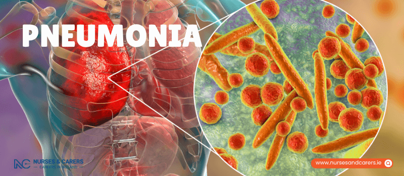
Pneumonia is an acute lower respiratory tract infection characterized by inflammation of the lung tissue, specifically the microscopic air sacs (alveoli). When infected, these air sacs fill with exudate, pus, and fluid, severely compromising pulmonary gas exchange. While the condition can be triggered by various pathogens, it is most commonly caused by a bacterial or viral infection.
Signs and Symptoms
Symptoms of pulmonary inflammation can manifest abruptly over a few hours or develop progressively over several days. The clinical presentation includes:
Productive Cough: A persistent cough that can be dry or produce thick yellow, green, or blood-flecked mucus (phlegm).
Dyspnoea: Shortness of breath or an accelerated respiratory rate.
Pyrexia: A high temperature or fever accompanied by sweating, chills, and rigors.
Thoracic Discomfort: Sharp chest pain that characteristically intensifies during deep inhalation or coughing (pleuritic pain).
Systemic Distress: Severe lethargy, generalized muscle body aches (myalgia), and a total loss of appetite.
Auscultatory Sounds: Wheezing or crackling during respiration. In infants, this often presents as distinct, rhythmic grunting noises with each breath.
Cognitive Shifts: Sudden confusion, disorientation, or delirium, particularly prevalent in elderly patients.
Pathological Causes and Subtypes
Bacterial Infections: The most prevalent and clinically severe cause of community-acquired pneumonia, frequently driven by Streptococcus pneumoniae.
Viral Infections: A primary driver of pediatric pneumonia. Viruses like influenza or SARS-CoV-2 can directly damage lung lining, and almost all critical or fatal complications of COVID-19 feature severe bilateral pneumonia.
Aspiration Pneumonia: Initiates when foreign material, such as vomit, food particles (e.g., a peanut), pool water, or toxic chemical smoke, is accidentally inhaled directly into the lungs, causing chemical irritation and secondary bacterial colonization.
Hospital-Acquired Pneumonia (HAP): Developed during an inpatient hospital stay. A subset includes ventilator-associated pneumonia, occurring when bacteria travel down a mechanical breathing tube (ventilator) directly into a compromised patient’s lungs.
Fungal Infections: Rare, opportunistic infections that primarily affect immunocompromised individuals.
Risk Stratification and Vulnerable Groups
While pneumonia can contract in any individual, certain populations carry an elevated risk of developing severe, life-threatening complications:
Infants, young children, and older adults due to developing or declining immune function.
Individuals with chronic co-morbidities, including heart failure, chronic obstructive pulmonary disease (COPD), asthma, liver cirrhosis, chronic kidney disease, or diabetes.
Patients living with advanced neurological conditions like Multiple Sclerosis (MS) that impair the swallowing reflex, increasing aspiration risks.
Individuals with severely suppressed immune systems (e.g., undergoing chemotherapy or living with HIV).
Active tobacco smokers, as nicotine paralyzes the lung's natural clearing mechanisms (cilia), allowing bacteria to settle.
Diagnostic Framework
To establish a definitive diagnosis and evaluate the severity of the infection, your GP will perform an immediate clinical workup:
Anatomical Auscultation: Listening to your chest and back using a stethoscope to identify diagnostic crackles, wheezing, or diminished breath sounds caused by fluid consolidation.
Biochemical Markers: Ordering blood tests to analyze full blood counts and measure C-Reactive Protein (CRP) to confirm systemic inflammation.
Radiological Imaging: Fast-tracking a chest X-ray to visually locate the exact site and extent of the fluid consolidation within the lung lobes.
Targeted Treatment Protocols
Therapeutic management is completely dictated by the underlying pathogen and the severity of the patient's baseline symptoms.
1. Home-Based Care (For Mild Pneumonia)
Mild cases can be safely resolved at home under targeted medical surveillance:
Antibiotic Therapy: If a bacterial strain is suspected, your GP will prescribe an immediate course of oral antibiotics. Note: Antibiotics are completely ineffective against viral pneumonia. However, viral cases are generally less severe unless secondary bacterial seeding occurs.
Supportive Measures: Prioritize absolute physical rest, maintain high fluid intake to thin respiratory secretions, and use paracetamol to regulate fever and manage thoracic pain.
2. Inpatient Hospitalization (For Severe Pneumonia)
If a patient displays clinical signs of respiratory failure or systemic instability, immediate hospital admission is mandatory. The inpatient protocol delivers:
Intravenous Therapies: Administering broad-spectrum antibiotics and hydration fluids directly into a vein through a cannula drip.
Oxygen Supplementation: Delivering humidified oxygen via a nasal cannula or face mask to maintain safe arterial oxygen saturation levels.
Strategic Prevention and Hygiene Control
1. Immunization Framework
If you fall into a high-risk category, you can secure public funding for preventative vaccines:
The Pneumococcal Vaccine: Directly targets the most common bacterial strains causing invasive lung disease. ⚠️Clinical Warning: This vaccine protects exclusively against specific pneumococcal bacteria; it does not prevent viral or alternative bacterial strains of pneumonia.
The Influenza Vaccine: Administered annually to prevent the primary seasonal viral infections that weaken lung tissue and lead to secondary pneumonia.
2. Cleanliness and Pulmonary Protection
Respiratory Etiquette: Always cover your mouth and nose with a disposable tissue when coughing or sneezing, and discard the tissue immediately before washing your hands thoroughly with antibacterial soap and water.
Lung Preservation: Actively quit smoking and significantly reduce alcohol consumption to restore the natural immune barriers of the upper respiratory tract.
Emergency Triage Action Plan
🚨 Emergency Medical Triage
Call 999, 112, or proceed to the nearest Emergency Department (ED) immediately if you or someone else develops:
Severe, unmanageable difficulty breathing or a feeling of suffocation.
A blue, grey, or pale discoloration of the lips, tongue, or nail beds (cyanosis, indicating profound oxygen deprivation).
An abrupt onset of acute confusion, severe drowsiness, or a total loss of consciousness.
⚠️ Urgent Clinical Triage
Contact your GP clinic immediately for an urgent, same-day consultation if you:
Feel progressively short of breath during minor daily movements.
Experience sharp, stabbing chest pain that noticeably intensifies every time you breathe in or out.
Have been managing a persistent, hacking cough that has lasted for 3 consecutive weeks or more.
Observe fresh blood mixed into your coughed-up phlegm (haemoptysis).
Treatment and Recovery
Diagnosing pneumonia can be clinically challenging because its primary indicators overlap significantly with more benign conditions like the common cold, acute bronchitis, or asthma flare-ups.
To isolate the condition, your GP will perform an immediate baseline workup:
Evaluating the timeline and severity of your physical symptoms.
Tracking your core temperature to detect pyrexia (fever).
Auscultation: Using a stethoscope to listen to your lungs. Fluid consolidation inside the lung sacs produces characteristic clicking, bubbling, or cracking sounds.
Note: If your symptoms are mild, physical exams are usually sufficient to begin care. If the clinician suspects moderate-to-severe disease, you will be fast-tracked for a confirmatory chest X-ray and inflammatory blood panels.
Home-Based Management (For Mild Pneumonia)
Most individuals diagnosed with mild pneumonia can recover completely at home using targeted rest and standard medical support.
Do
Complete Your Antibiotic Course: If your GP suspects a bacterial infection, they will prescribe an immediate course of oral antibiotics. You must finish the entire packet exactly as directed, even if you begin to feel better within a few days.
Maintain High Hydration: Drink large volumes of water and clear fluids daily. Excellent hydration physically thins thick respiratory secretions, making them significantly easier to cough up.
Prioritize Rest: Remain in bed or rest continuously until your baseline energy returns and your temperature stabilizes.
Manage Symptoms Safely: Use oral paracetamol or ibuprofen to safely lower a high temperature and dull pleuritic chest pain. Sip warm honey and lemon water to naturally soothe a raw, irritated throat.
Isolate Wisely: It is completely safe to be around healthy family members at home. However, you must strictly avoid close contact with anyone who is elderly, newborn, or living with a compromised immune system.
Don't
Do Not Smoke: Completely avoid smoking tobacco or vaping, as nicotine and hot vapors paralyze the lung's natural clearing mechanisms (cilia) and severely delay tissue healing.
Avoid Over-the-Counter Cough Suppressants: Do not take commercial cough medicines. Coughing is your body's vital defense mechanism required to mechanically expel infected pus and fluid from your lung tissue. Suppressing the cough reflex can trap the infection and worsen the condition.
Inpatient Hospitalization (For Severe Pneumonia)
If your symptoms are severe, or if an initial course of home antibiotics fails to halt the infection, immediate inpatient admission is required. Hospital teams will deliver:
Intravenous (IV) Therapies: Administering broad-spectrum antibiotics and replacement fluids directly into your bloodstream via an intravenous drip to bypass the digestive tract.
Oxygen Supplementation: Delivering humidified oxygen through a nasal cannula or face mask to maintain your arterial oxygen saturation at a safe clinical level.
Managing Aspiration via Bronchoscopy
If your condition is classified as aspiration pneumonia—meaning the lung inflammation was triggered by accidentally inhaling a foreign object (such as a piece of food or a peanut)—your medical team must clear the mechanical blockage.
The Procedure: A specialist performs a bronchoscopy, passing a thin, flexible tube equipped with a miniature camera and micro-instruments through your nose or mouth down into your airways to locate and safely extract the obstructing object.
The Recovery Timeline and Post-Care Staging
Complete recovery from a lower respiratory tract infection takes time, and healing milestones occur progressively over several weeks:
Weeks 2 to 3: It is completely standard to manage a lingering, residual cough for 2 to 3 weeks after completing your full course of antibiotics.
Month 1 and Beyond: Systemic fatigue, physical weakness, and low energy levels frequently persist for a month or more as your immune system repairs the cellular damage to your lungs.
The 6-Week Review: You will attend a mandatory follow-up appointment with your GP approximately 6 weeks after initiating your antibiotic treatment to verify full clinical resolution.
Advanced Post-Recovery Screening
Your GP will schedule a follow-up chest X-ray at your 6-week review to ensure your lung fields are entirely clear if:
Your physical symptoms have failed to improve or have actively returned.
You are a current or historical tobacco smoker.
You are over the age of 50.
Preventative Care Step: Once your lungs have fully recovered, your GP will evaluate your eligibility for the annual influenza vaccine and the one-off pneumococcal vaccine to protect you against future pulmonary infections.
Clinical Triage and Action Plan
🩺 Non-Urgent Medical Tracking
Contact your GP clinic for advice if:
Your fever or respiratory symptoms show zero improvement within 3 days (72 hours) of initiating your prescription antibiotics. This failure to respond can indicate that the underlying pathogen is a virus (which does not respond to antibiotics) or that the bacteria are resistant to the specific drug prescribed, requiring an immediate medication switch.
Urgent Same-Day Clinical Triage
Contact your GP clinic immediately or proceed to an urgent care unit if you develop:
Progressive shortness of breath during minor movements or while at rest.
Sharp, stabbing chest pain that characteristically intensifies every time you breathe in or out.
A hacking, persistent cough that has lasted for 3 consecutive weeks or longer.
Fresh blood mixed into your coughed-up phlegm (haemoptysis).
Immediate Emergency Response
Call 999, 112, or proceed to your nearest hospital Emergency Department (ED) immediately if you or someone else exhibits:
Acute, unmanageable difficulty breathing, gasping for air, or an inability to speak in full sentences.
A blue, grey, or pale discoloration of the lips, tongue, or nail beds (cyanosis, indicating critical oxygen deprivation).
A sudden onset of severe confusion, profound drowsiness, or a total loss of consciousness.
Pneumonia in Babies and Children
Pneumonia in pediatric patients is an acute inflammation of the lung tissue that frequently manifests as a secondary complication immediately after a child has been fighting off a primary upper respiratory tract infection, such as a common cold or the seasonal flu. Because a child’s respiratory system is small and can deteriorate quickly, you must seek prompt medical evaluation if you suspect they are developing lung inflammation.
Signs and Symptoms in Children
A child's respiratory symptoms can develop abruptly over a few hours or manifest progressively over several days. Key systemic and behavioral indicators include:
A high temperature or sudden fever.
Chronic lethargy, sleeping significantly more than usual, or extreme weakness.
Increased irritability, persistent crying, and an inability to be easily soothed.
A hacking cough that can be dry or produce thick yellow, green, or brown mucus (phlegm).
Localized chest pain or abdominal discomfort caused by coughing strains.
A sharp decline in fluid intake, or a total refusal to bottle-feed, breastfeed, or drink water.
Mechanical Signs of Respiratory Distress
Children use auxiliary muscle groups to compensate when their lungs are inflamed. Actively inspect your child's bare chest and face for these physical indicators of labored breathing:
Nasal Flaring: The nostrils visibly widen or flare outward with every single inhalation.
Abdominal Exertion: The child's tummy muscles contract hard and push out aggressively to force air in and out.
Intercostal Retractions: The skin between, below, or around the ribs distinctly "sucks in" with each breath.
Auscultatory Noises: High-pitched wheezing sounds during breathing, or rhythmic, short grunting noises made by babies as they exhale to try to keep their airways open.
The Diagnostic Framework
Your GP can routinely diagnose pediatric pneumonia in the clinic by performing a thorough physical examination and using a stethoscope to listen to your child's chest and back for diagnostic fluid crackles. If the presentation is complex or severe, the doctor will refer your child for hospital-based tracking tests:
Confirmatory Chest X-ray: To pinpoint the exact location of the fluid consolidation in the lungs.
Biochemical Blood Tests: To evaluate white blood cell responses and track inflammatory markers.
Caregiver Note: You have the absolute right to remain directly alongside your child to offer comfort and safety during all imaging and blood collection procedures.
Pediatric Treatment Protocols
1. Frontline Medical Options
Bacterial Infections: Your GP will prescribe a targeted course of pediatric oral liquid antibiotics. This medication must be completed entirely according to the exact schedule, even if the child's fever drops and they appear to recover early.
Viral Infections: Antibiotics are completely ineffective against viral pneumonia. Viral strains are highly common in young children and typically run a milder course, requiring supportive home care while the virus clears naturally.
2. Criteria for Inpatient Hospitalization
If your child is exceptionally ill, completely refusing fluids, or persistently vomiting and unable to retain oral medications, they must be admitted to the hospital. Inpatient pediatric units will deliver:
Intravenous Access: Administering broad-spectrum antibiotics and rehydration fluids directly into a vein via an intravenous cannula drip.
Respiratory Support: Administering humidified oxygen through a specialized pediatric face mask or lightweight nasal prongs to ease their breathing.
Home Care and Recovery Guidance
Do
Prioritize Absolute Rest: Keep your child calm, quiet, and resting at home until they are entirely free of fever and their energy levels normalize.
Manage Pain and Fever Wisely: Administer specialized pediatric liquid formulations of paracetamol or ibuprofen to lower a high temperature and reduce chest soreness.
⚠️ Infant Safety Restriction: Never administer ibuprofen to an infant under the age of 3 months unless explicitly directed by a medical doctor.
Increase Fluid Frequency: If you are breastfeeding or bottle-feeding a baby, offer smaller feeds significantly more often throughout the day to prevent dehydration without overwhelming their stomach. Encourage older children to take frequent sips of water.
Maintain Immunization Schedules: Ensure your child is fully up-to-date with their primary childhood vaccination schedule, as vaccines against Hib, pertussis, and pneumococcal bacteria directly protect them against the most lethal strains of pneumonia.
Enforce Hygiene Boundaries: Teach your child excellent handwashing habits and instruct them never to share drinking cups, water bottles, or cutlery with peers.
Don't
Enforce a Strict Smoke-Free Zone: Never smoke or allow anyone else to smoke tobacco or vape around your child. Second-hand smoke paralyzes the lung's protective defenses, severely worsens respiratory inflammation, and delays tissue recovery.
Avoid Pediatric Cough Suppressants: Do not give over-the-counter cough medicines to any child under the age of 6 (unless specifically prescribed by your GP). Coughing is a critical mechanical reflex required to clear infected phlegm from the lungs; blocking it traps the infection and can cause respiratory complications.
Clinical Triage Action Plan
Immediate Emergency Response
Call 999, 112, or proceed to your nearest hospital Emergency Department (ED) immediately if your child exhibits:
High-volume gasping for air, severe difficulty breathing, or breathing that is abnormally rapid and shallow.
A blue, grey, or pale discoloration of the lips, tongue, or skin surrounding the mouth (indicating critical oxygen deprivation).
Extreme lethargy where the child is floppy, unresponsive, or exceptionally difficult to wake up.
Urgent Medical Action Required
Contact your GP clinic immediately for an urgent, same-day appointment if:
You observe any of the baseline signs of pediatric pneumonia, including nasal flaring, grunting, or rib retractions.
Your child’s symptoms or fever fail to show visible signs of improvement within 3 days (72 hours) of starting their prescription antibiotics.
Disclaimer
The information provided here is for educational and informational purposes only. It does not constitute medical advice, diagnosis, or treatment. Always consult a qualified healthcare professional or your GP for specific medical concerns, diagnoses, or health advice. Never disregard professional medical advice or delay seeking it because of information you have read here.