
Pleurisy
17 Sept 2026 · 4 min read
On this page19 sections
Overview
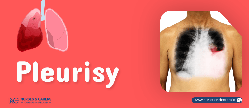
Pleurisy is the inflammation of the pleura—the dual-layered serous membrane that lines the thoracic cavity and envelops the lungs. While the condition is typically straightforward to treat and resolves within a few days, it can serve as a clinical indicator of a more severe underlying pulmonary or systemic disorder, such as pneumonia.
Symptoms of Pleural Inflammation
The clinical presentation of pleurisy is characterized by sharp, localized thoracic discomfort directly tied to respiratory movement. Common symptoms include:
Pleuritic Chest Pain: A sharp, stabbing sensation in the chest that intensifies acutely during inhalation, coughing, sneezing, or physical movement.
Referred Discomfort: Pain radiating upward into the shoulder region.
Respiratory Compensation: Discomfort that noticeably diminishes when taking shallow, rapid breaths.
Secondary Signs: Shortness of breath (dyspnoea) and a dry, non-productive cough.
Pathological Causes
The most common cause of pleurisy is a viral respiratory tract infection, such as influenza. When driven by a virus, the inflammation is usually self-limiting.
2. Secondary and Complex Triggers
Bacterial Infections: Lower respiratory tract infections, including bacterial pneumonia or tuberculosis.
Pulmonary Embolism (PE): A life-threatening blood clot that occludes an artery in the lungs.
Mechanical Trauma: Blunt force injuries to the chest wall, such as bruised or fractured ribs, which directly irritate the adjacent pleura.
Malignancy: Primary lung cancer or metastatic pleural disease.
Autoimmune Disorders: Chronic systemic inflammatory conditions like lupus or rheumatoid arthritis.
Respiratory Syndromes (including COVID-19): While viruses like SARS-CoV-2 may not always directly infect the pleura, they cause severe secondary complications like pneumonia that frequently lead to pleurisy.
The Clinical Diagnostic Pathway
Primary Care Assessment
During a physical examination, your GP will use a stethoscope to listen to your lungs. Inflamed pleural layers lose their smooth lubrication and rub together during respiration, creating a characteristic dry, grating, or crunching sound known as a pleural friction rub.
Advanced Diagnostic Workup
To isolate the exact cause of the inflammation, your care team may coordinate several targeted tests:
Biochemical Blood Panels: To check for elevated inflammatory markers or autoimmune antibodies.
Chest X-ray: To evaluate the lung structures and rule out bone fractures or pneumonia.
Ultrasound or CT Scans: High-resolution cross-sectional imaging to visualize tissue changes and check for fluid.
Pleural Biopsy: Excising a micro-sample of the pleural tissue for laboratory microscopic analysis.
Targeted Treatment Protocols
When addressed promptly, pleurisy typically resolves completely without inflicting any permanent or lasting damage to the lung tissues. Clinical management focuses on pain eradication and treating the underlying disease.
1. Managing Pleuritic Pain
First-Line Anti-inflammatories: Non-steroidal anti-inflammatory drugs (NSAIDs) like ibuprofen are the gold standard for pleurisy because they directly target and reduce the tissue inflammation driving the pain. If NSAIDs are contraindicated for your health profile, your GP will prescribe an alternative analgesic.
Postural Offloading: When resting, try lying directly on the painful side of your chest. This physically restricts the expansion of that specific lung, minimizing the friction between the inflamed membranes and lowering pain.
2. Eradicating the Root Cause
Viral Protocols: Require zero targeted medications; treatment relies entirely on rest, hydration, and symptom relief while the virus clears naturally.
Bacterial Protocols: Require a strict course of prescription oral or intravenous antibiotics.
Note: If you have pre-existing health conditions or exhibit severe respiratory distress, you may require inpatient hospital admission.
3. Managing Secondary Pleural Effusion
In some cases, inflamed tissue leaks excess fluid into the space between the membranes, a complication known as pleural effusion. While the fluid temporarily separates the membranes and can lessen the sharp pain, it physically compresses the lung, leading to worsening shortness of breath.
Surgical Drainage (Thoracentesis): If the fluid fails to reabsorb naturally or severely compromises your breathing, it must be mechanically removed. Performed under local or general anaesthesia, a specialist inserts a hollow needle or a chest tube through the ribs to drain the trapped fluid. If high volumes of fluid require slow drainage, a brief hospital stay of a few days is mandatory.
Clinical Triage and Action Plan
⚠️ Urgent GP Review Required
Contact your GP clinic immediately for an urgent appointment if:
You experience sharp, stabbing chest pains specifically when breathing in, coughing, or sneezing.
Immediate Emergency Care
Call 999, 112, or go to the nearest Emergency Department (ED) immediately if:
Your chest pain is sudden, crushing, severe, or unmanageable.
Your chest pain is accompanied by coughing up blood (haemoptysis), profound nausea, or profuse sweating.
The thoracic pain suddenly intensifies and spreads down across your abdomen.
Your chest pain briefly improves or disappears before returning with significantly worse intensity.
Disclaimer
The information provided here is for educational and informational purposes only. It does not constitute medical advice, diagnosis, or treatment. Always consult a qualified healthcare professional or your GP for specific medical concerns, diagnoses, or health advice. Never disregard professional medical advice or delay seeking it because of information you have read here.