
Echocardiogram (Echo)
9 Sept 2026 · 3 min read
On this page8 sections
An echocardiogram (echo) is a safe, painless hospital scan that uses ultrasound waves to create detailed images of the heart and its surrounding blood vessels.
Unlike an electrocardiogram (ECG)—which only measures your heart's electrical activity and rhythm—an echo physical evaluates the heart's actual structure, muscle condition, and blood flow.
Why It Is Used
Cardiologists use an echocardiogram to determine the best treatment plan for various conditions by detecting:
Heart Failure: Tracking how efficiently the heart pumps blood.
Heart Attack Damage: Assessing areas where blood supply was previously blocked.
Valve Problems: Checking the chambers that regulate internal blood flow.
Cardiomyopathy: Identifying thickened or enlarged heart walls.
Endocarditis: Spotting infections on the heart valves.
Congenital Heart Disease: Diagnosing heart defects present from birth.
The Standard Test: Transthoracic Echocardiogram (TTE)
The TTE is the most common type of echo. It requires no advanced preparation and takes about 25 minutes.
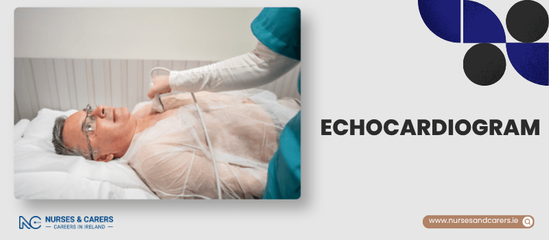
Setup: You remove top-half clothing and lie on your left side.
Monitoring: Sticky sensors (electrodes) are attached to your chest to track your rhythm.
Scanning: A specialist applies cold lubricating gel and glides a handheld ultrasound probe across your chest.
Experience: You may hear a "swishing" sound, which is simply the audio of your blood flowing.
Recovery: You can go home immediately. Skin may feel slightly tender when the sticky patches are removed.
Other Types of Echocardiograms
If your doctor needs more detailed images or wants to see your heart under physical strain, they may request an alternative method:
1. Transoesophageal Echocardiogram (TOE)
Purpose: Provides highly detailed images, often to plan for an upcoming heart surgery.
Process: A doctor numbs your throat with spray, gives you a relaxing sedative, and passes a thin probe down your food pipe.
Preparation: You must fast (no food or drink) for several hours beforehand.
Recovery: Your throat may feel sore. You cannot eat or drink for 2 hours after the test until the numbness wears off. Because of the sedative, you must arrange a ride home and avoid driving, alcohol, or heavy machinery until the next day.
2. Stress Echocardiogram
Purpose: Used if your heart symptoms are triggered specifically by physical exertion.
Process: The scan is performed during or right after you use a treadmill/exercise bike, or after receiving a medication injection that mimics exercise.
Risks: You might experience temporary nausea, dizziness, or chest pain. The medical team monitors you continuously and will stop the test immediately if any irregular rhythms occur.
3. Contrast Echocardiogram
Purpose: Enhances image clarity to see blood flow patterns perfectly.
Process: A harmless contrast dye is injected into your bloodstream before the ultrasound.
Risks: Safe for most, though minor allergic reactions like itchy skin can occasionally occur.
Getting Your Results
You will not receive your results immediately on the day of the scan. The imaging specialist will analyze the ultrasound data and securely transfer the report to your referring doctor, who will discuss the findings with you at your next scheduled appointment.
Disclaimer
The information provided here is for educational and informational purposes only. It does not constitute medical advice, diagnosis, or treatment. Always consult a qualified healthcare professional or your GP for specific medical concerns, diagnoses, or health advice. Never disregard professional medical advice or delay seeking it because of information you have read here.