
Electrocardiogram (ECG)
9 Sept 2026 · 3 min read
On this page6 sections
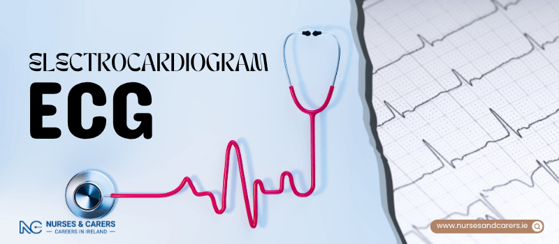
An electrocardiogram (ECG) is a quick, safe, and painless medical test used to evaluate your heart’s rhythm and electrical activity. Every time your heart beats, it generates tiny electrical signals; an ECG detects these impulses using sensors attached to your skin and records them onto a machine.
An ECG is distinctly different from an echocardiogram (echo), which is an ultrasound scan that captures physical images of the heart's structure.
Why an ECG is Performed
A doctor or cardiologist may request an ECG to investigate unexplained symptoms, including:
Chest pain or a heavy, crushing sensation.
Palpitations, where the heart feels like it is fluttering, pounding, or skipping a beat.
Shortness of breath or sudden, unexplained dizziness.
The test helps medical professionals diagnose and monitor critical heart conditions such as:
Arrhythmias: Irregular, abnormally fast, or unusually slow heartbeats.
Coronary Heart Disease: Reduced blood flow to the heart muscle due to narrowed arteries.
Heart Attacks: Acute blockages preventing blood supply to parts of the heart.
Cardiomyopathy: Structural changes where the heart walls become abnormally thick or enlarged.
Medication Tracking: Monitoring patients taking specific prescription drugs that can alter heart function.
The Three Main Types of ECG
The specific test your doctor selects will depend on your symptoms and what triggers them:
Resting ECG: The standard procedure performed while you are lying down completely still.
Stress or Exercise ECG: Conducted while you walk on a treadmill or pedal an exercise bike. It is used if your chest symptoms or irregularities are specifically triggered by physical exertion.
Ambulatory ECG (Holter Monitor): You wear a small, portable recording device home for 24 hours or longer. This is ideal for tracking unpredictable symptoms that happen in short, random episodes.
How the Procedure is Done
There is no advanced preparation required for a standard ECG; you can eat and drink normally beforehand.
Setup: You will be asked to remove your upper-body clothing. A nurse or technician may clean or shave small patches of hair from your chest to ensure a clean signal.
Electrodes: Small, sticky sensors called electrodes are attached to your chest, arms, and legs. These sensors are linked via wires to a recording machine.
The Scan: Once the wires are connected, your chest is covered. You lie still for a few minutes while the machine records.
Completion: You can leave the clinic immediately after the electrodes are removed.
Risks and Side Effects
An ECG is highly safe. The machine does not send any electricity into your body; it only listens to your heart's natural output.
Skin Reactions: You might feel a slight tugging sensation—similar to removing a plaster—when the sticky pads are peeled off. A mild, temporary rash may occur at the sensor sites.
Exercise ECG Safety: During a stress ECG, trained medical staff monitor you continuously and will stop the machine immediately if you report feeling unwell, dizzy, or experience chest pain.
Getting Your Results
The ECG machine translates your heart's electrical patterns into a wave-like graph displayed on a screen or printed on paper. For ambulatory monitors, the data is saved electronically for analysis.
Because a specialist or cardiologist must carefully review the graphs, your results are rarely available immediately. Your doctor will schedule a follow-up appointment a few days later to discuss the findings and determine if further diagnostic tests are necessary.
Disclaimer
The information provided here is for educational and informational purposes only. It does not constitute medical advice, diagnosis, or treatment. Always consult a qualified healthcare professional or your GP for specific medical concerns, diagnoses, or health advice. Never disregard professional medical advice or delay seeking it because of information you have read here.