
Epidermolysis Bullosa (EB)
10 Sept 2026 · 15 min read
On this page36 sections
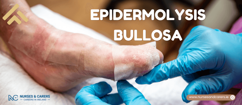
Epidermolysis Bullosa (EB) refers to a group of rare, inherited dermatological conditions characterized by extreme fragility of the skin and mucous membranes. Individuals with EB experience painful blistering, erosions, and scarring in response to minor friction, rubbing, or minor trauma.
Common Symptoms
While clinical presentation varies by type, core symptoms across all forms of EB include:
Fragile Skin: Stripping, blistering, and tearing from minimal contact.
Localized Blistering: Frequent blisters developing on high-friction areas, particularly the hands and the soles of the feet.
Dermatological Changes: Thickening of the skin and nails over time, hyperpigmentation or hypopigmentation, and extensive cutaneous scarring.
The Four Main Types of EB
EB is categorized into four primary types based on the specific layer of skin where blistering occurs. Each type contains multiple subtypes with varying degrees of severity:
Epidermolysis Bullosa Simplex (EBS): The most common form, where blistering typically occurs in the outermost layer of skin (epidermis).
Dystrophic Epidermolysis Bullosa (DEB): Characterized by blisters forming below the basement membrane, often leading to severe scarring.
Junctional Epidermolysis Bullosa (JEB): A severe form where blisters form within the basement membrane zone.
Kindler Epidermolysis Bullosa (KEB): A rare variant where blistering can occur across multiple skin layers, accompanied by photosensitivity.
Diagnosis and Prenatal Testing
Postnatal Diagnosis
EB is predominantly diagnosed during infancy or early childhood, though milder forms may evade detection until adulthood. If a general practitioner (GP) suspects EB, they will issue an urgent referral to a pediatric or general dermatologist. The specialist will confirm the diagnosis, identify the specific EB type, and establish a multidisciplinary treatment plan using:
Genetic Testing: Analysing a standard blood sample to map specific genetic variants.
Skin Biopsy: Taking a small sample of affected skin for specialized diagnostic laboratory evaluation.
Prenatal Diagnostic Testing
Testing can be performed on an unborn baby after the 11th week of pregnancy via amniocentesis or chorionic villus sampling (CVS). These tests are typically offered if:
Either parent is a confirmed carrier of the genetic variant linked to EB.
There is a documented family history indicating a high risk of conceiving a child with a severe form of EB.
Note: If a prenatal test confirms the diagnosis, comprehensive clinical counselling and family support options are provided.
Causes and Genetic Transmission
EB is caused by mutations in genes responsible for producing the structural proteins that bind the layers of the skin together. Without these anchoring proteins, the skin layers easily separate. The genetic alteration can be inherited in a few ways:
Autosomal Dominant: Inherited from a single parent who actively has the condition.
Autosomal Recessive: Inherited from both parents, who carry the recessive gene variant but do not display symptoms themselves.
De Novo Mutation: Spontaneous genetic changes occurring purely by chance, with no parental history of the variant.
Treatment and Care Management
There is currently no cure for EB. Medical management is supportive and focused on preserving skin integrity, relieving chronic pain, accelerating wound healing, and preventing secondary infections or disabling complications.
A specialized, multidisciplinary medical team works closely with the family to navigate daily wound care, bandage selections, and lifestyle adjustments.
Psychological Support and Community Resources
Receiving an EB diagnosis can cause profound emotional distress, including anxiety, grief, and fear. Accessing specialized support networks is critical for both the individual and their caregivers.
Patient and Family Support Networks
DEBRA Ireland (debra.ie) – The dedicated national charity providing support, advocacy, and research funding for EB.
Irish Skin Foundation (irishskin.ie) – Offering targeted educational resources for diverse skin conditions.
Children's Health Ireland (childrenshealthireland.ie) – Providing pediatric clinical excellence and family support frameworks.
Carer Welfare
Caring for a child with a complex, medically demanding condition like EB involves significant physical and emotional labor. Caregivers are strongly encouraged to utilize dedicated respite care services, state benefits, and psychological support networks.
Clinical Overview of Symptoms
The symptoms of Epidermolysis Bullosa (EB) vary significantly based on the genetic type and subtype diagnosed. While some forms present as localized, manageable discomfort, other variants involve widespread systemic complications that can be life-threatening, particularly in infancy.
1. Epidermolysis Bullosa Simplex (EBS)
EBS presents on a spectrum from localized blistering to severe, widespread cutaneous involvement. Symptoms typically worsen during hot weather or periods of increased sweating.
Localised EBS (Most Common)
Presentation: Painful blistering primary confined to high-friction areas like the palms of the hands and soles of the feet.
Triggers: Mild to moderate physical exertion, such as walking, running, or cycling (which can cause friction blisters on the buttocks and inner thighs).
Prognosis: Blisters typically heal without permanent scarring.
Adulthood Manifestations: Symptoms can emerge from early childhood into adulthood. Over time, adults often develop hyperkeratosis (thickening of the skin) on the palms and soles, alongside thickened fingernails and toenails.
Intermediate EBS
Presentation: Generalized blistering can appear anywhere on the body in response to minor trauma or friction.
Onset: Birth or early childhood.
Key Symptoms: Mild mucosal blistering inside the mouth, localized scarring, milia (tiny white skin spots), and progressive thickening of the nails, palms, and soles.
Generalised Severe EBS
Presentation: Extreme, widespread blistering evident immediately at birth. Infants can develop up to 200 new blisters daily.
Complications:
Infection & Feeding Issues: Extensive blistering compromises the skin barrier, increasing infection risks and disrupting feeding patterns, which can cause developmental delays.
Mobility: Severe plantar blisters can delay a toddler's ability to walk.
Nails: Complete loss or severe thickening of fingernails and toenails.
Prognosis: Cutaneous blistering gradually settles with age, though adults retain heavily thickened skin on their palms and soles, which can permanently impair mobility.
2. Junctional EB (JEB)
JEB impacts both the external skin and internal mucous membranes (soft tissue linings).
Intermediate JEB
Onset: Birth or early infancy, occasionally improving slightly with age.
Key Symptoms: Widespread blistering, slow-healing ulcerations (particularly on the lower legs), nail loss, and large, irregular moles.
Alopecia: Scalp blistering can lead to permanent scarring and hair loss.
Systemic Impact: Enamel hypoplasia (damaged tooth enamel), severe dental decay, and painful oral blisters that restrict eating. Scarring may also affect the eyes and urethra.
Oncology Risk: Adults face a significantly elevated risk of developing skin cancer. Bi-annual (twice-yearly) dermatologist check-ups are highly recommended.
Severe JEB
Presentation: Extensive, devastating blistering and ulceration across the skin, respiratory tract, and internal organs.
Anatomical Sites: Frequently targets the mouth, throat, nose, eyes, neck, digits, anus, and genitalia.
Secondary Complications: Severe anemia, rapid dehydration, profound malnutrition, stunted growth, acute respiratory distress, and life-threatening sepsis.
Prognosis: Due to these severe systemic complications, many infants with severe JEB do not survive past their first few months of life.
3. Dystrophic EB (DEB)
DEB is characterized by deep tissue blistering that leads to prominent scarring and structural changes.
Dominant DEB
Presentation: Mild to moderate blistering localized primarily to the extremities (hands, feet, arms, and legs) that resolves with scarring and milia.
Dermatological Signs: Dystrophic (abnormally shaped) or entirely absent nails. Oral involvement can make eating and dental hygiene highly painful.
Onset: Birth through late childhood. In mild cases, missing or misshapen nails may be the only visible clinical sign.
Recessive DEB (Severe Variant)
Cutaneous Symptoms: Extensive, chronic blistering and ulceration present at birth, often leaving large patches of missing skin.
Pseudosindactilia (Mitten Deformity): Repeated, severe scarring on the hands and feet can cause the skin between the fingers and toes to fuse together, alongside complete nail loss. Scarring on the scalp leads to permanent patchy alopecia.
Mucosal & Systemic Involvement: Deep blistering occurs throughout the mouth, esophagus, and digestive tract. Microstomia (narrowing of the mouth opening) and esophageal strictures cause severe pain when chewing or swallowing.
Secondary Complications: Chronic anemia, severe malnutrition, marked growth failure, and painful ocular scarring that leads to vision impairment.
Oncology Risk: This cohort carries an exceptionally high risk of developing aggressive skin cancers. Mandatory clinical skin checks with a dermatologist are required at least twice a year.
4. Kindler EB (KEB / Kindler Syndrome)
KEB is an exceedingly rare, multi-system variant of EB.
Cutaneous Symptoms: Widespread blistering begins at birth but progressively decreases with age. It is uniquely characterized by severe photosensitivity (extreme vulnerability to sun damage).
Dermatological Changes: Progressive poikiloderma (skin discoloration), thinning and wrinkling of the skin on the hands, feet, and neck, hyperkeratosis of the palms and soles, and the complete loss of epidermal ridges (disappearance of fingerprints).
Internal Involvement: Chronic inflammation, blistering, and scarring can severely damage the teeth, gums, esophagus, eyelids, and eyes.
Oncology Risk: Similar to JEB and Recessive DEB, individuals with KEB have an elevated risk of skin cancer, necessitating dermatological screenings at least twice a year.
Treatment Goals and Clinical Framework
There is currently no cure for Epidermolysis Bullosa (EB). The primary objectives of clinical management are to alleviate painful symptoms, preserve skin integrity, accelerate wound healing, and prevent secondary complications like systemic infections.
Specialist Multidisciplinary Care
Upon diagnosis, patients are referred to a specialist tertiary care centre to establish a comprehensive, individualized treatment plan. While severe forms require ongoing management at these specialized hubs, patients with stable or milder symptoms may eventually transition to shared care closer to home.
Because EB impacts multiple organ systems, care is delivered by a Multidisciplinary Team (MDT), which typically includes:
Dermatologist: Oversees specialized skin assessments and advanced wound care protocols.
Specialist Nurse: Coordinates communication between the family and the medical team.
Dietitian: Optimizes nutritional intake to counter malabsorption and support wound healing.
Physiotherapist: Maintains joint mobility, manages scar tissue, and preserves physical function.
Dentist: Provides specialized, low-friction oral hygiene strategies.
Play Specialist: Employs therapeutic activities to support the child's physical and psychological well-being.
Daily Preventative Strategies
To minimize friction-induced skin damage while encouraging participation in normal childhood activities, implement the following daily adjustments:
Skin Protection: Apply advanced protective dressings and targeted foam padding to high-friction anatomical zones.
Thermoregulation: Keep the patient as cool as possible during warm weather, as increased sweating exacerbates blistering.
Clothing: Select loose-fitting garments made exclusively from soft, natural fabrics (such as 100% cotton). Avoid clothing with tight waistbands, coarse seams, or tags that rub against the skin.
Footwear: Choose comfortable, well-fitting shoes that feature completely smooth interior seams.
Advanced Skin and Wound Care
Proper wound management is essential to minimize pain and prevent extensive tissue loss. Never use standard adhesive bandages or regular sticking plasters, as removing them will strip away the fragile epidermis.
Blister Lancing: New blisters should be promptly punctured (lanced) using a sterile needle (supplied by a GP or specialist center). Lancing prevents fluid accumulation from expanding the blister and creating a larger, more painful wound.
Technique: Pierce the base of the blister to drain the fluid, but leave the overlying skin intact. This top layer acts as a natural biological barrier to protect the raw tissue underneath.
Dressing Application: Cover open wounds with specialized, non-adherent (non-stick) dressings designed for fragile skin. Secure these dressings in place using soft cotton bandages, tubular retention bandages, or specialized socks rather than medical tape.
Moxturization: Apply prescribed emollient creams regularly to support skin elasticity, as directed by the care team.
Monitoring and Managing Secondary Infections
Open lesions and raw skin patches are highly susceptible to bacterial colonization. Because untreated skin infections can rapidly progress to life-threatening systemic sepsis—particularly in severe forms of EB—prompt medical intervention is crucial.
Clinical Red Flags for Infection
Contact your GP or specialist care team immediately if you observe any of the following signs:
Increased heat, swelling, or spreading redness around a wound (note: erythema may be more subtle or present differently on darker skin tones).
The presence of purulent discharge (pus) or excessive watery fluid leaking from the wound.
Yellowish crusting or scabbing on the wound surface, or a wound that fails to progress toward healing.
Red streaks tracking away from a blister site, or a sudden, dense cluster of new blisters.
A systemic fever or body temperature reaching 38°C (100.4°F) or higher.
Medical Interventions for Infection
Depending on the severity, infections are managed using targeted antiseptic creams, topical antibiotic lotions, oral antibiotic tablets, or specialized antimicrobial wound dressings.
Symptom-Specific Clinical Support
1. Pain Management
Chronic and acute pain from dressings and wounds can severely restrict basic mobility.
Mild Forms: Managed using standard over-the-counter analgesics, such as paracetamol or ibuprofen.
Severe/Chronic Pain: Requires prescription-strength analgesics, including specialized opioids like morphine.
Crucial Safety Warning: Never administer aspirin to children or adolescents under the age of 16. Aspirin use in this age group is linked to a rare but life-threatening condition known as Reye’s syndrome.
2. Advanced Nutrition and Gastrointestinal Care
Epidermolysis bullosa increases metabolic demands due to continuous wound healing, placing patients at a high risk for malnutrition and anemia.
Oral Blistering: When oral mucosa lesions make feeding difficult for infants, specialized syringes or specialized soft-tipped eyedroppers may be required.
Dietary Modifications: For older children, incorporate soft, high-calorie, and high-protein foods. Serve all meals at room temperature or lukewarm, as hot foods can cause instant oral blistering.
Supplements & Testing: Regular blood panels are conducted to check for micro-nutrient deficiencies. Based on the results, clinicians may prescribe high-calorie nutritional shakes, or iron, zinc, and vitamin supplements.
Managing Constipation: Blistering around the anus makes defecation highly painful, which frequently leads to stool withholding and chronic constipation. This issue is often compounded as a side effect of opioid pain medications. Manage this early using prescribed fiber supplements or osmotic laxatives.
3. Dental, Nail, and Ocular Care
Dental Care: Maintain rigorous oral hygiene using an ultra-soft toothbrush and a fluoride mouthwash to counter the high risk of tooth decay caused by enamel defects.
Nail Care: Sub-ungual blisters can cause fingernails and toenails to become severely thickened and distorted. Apply prescribed keratolytics (nail-softening creams) to make trimming safer and easier.
Eye Care: Severe variants can cause painful corneal abrasions and ocular blistering. The MDT will routinely monitor eye health and may prescribe lubricating eyedrops or ophthalmic ointments to preserve moisture and protect the ocular surface.
Surgical Interventions
Patients with severe forms of EB may require targeted surgical procedures to correct structural complications arising from chronic scar tissue.
Hand and Foot Surgery (Separation of Digits): Repeated blistering and scarring can cause pseudo-syndactyly (where fingers and toes fuse together into a "mitten" deformity). Surgery is performed to separate the digits and restore manual dexterity.
Esophageal Dilation: If chronic scarring narrows the food pipe (esophagus) and causes dysphagia, a surgeon can perform an endoscopic dilation. A small balloon is inserted into the esophagus and carefully inflated to widen the stricture.
Gastrostomy Tube Insertion (G-Tube Surgery): If severe oral or esophageal involvement prevents the patient from consuming enough nutrients to sustain growth, a feeding tube is surgically placed directly into the stomach to ensure safe, adequate caloric intake.
Clinical Trials
Clinical trials represent a vital avenue for accessing emerging, next-generation therapies for EB. Speak directly with your specialist MDT to evaluate whether your child meets the eligibility criteria for current research studies. While clinical trials offer the potential benefit of novel treatments, they carry the inherent risk that the experimental therapy may not outperform standard care protocols.
Disclaimer
The information provided here is for educational and informational purposes only. It does not constitute medical advice, diagnosis, or treatment. Always consult a qualified healthcare professional or your GP for specific medical concerns, diagnoses, or health advice. Never disregard professional medical advice or delay seeking it because of information you have read here.