
Pacemaker
17 Sept 2026 · 19 min read
On this page11 sections
When You Might Need One
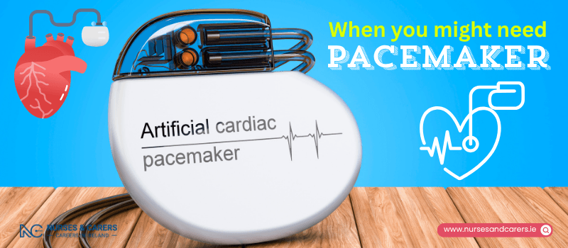
A pacemaker is a small electronic device implanted under the skin to regulate an abnormal heart rhythm (arrhythmia), most commonly when the heart beats too slowly. Pacemaker implantation is a highly routine cardiovascular procedure that significantly enhances quality of life and can be lifesaving.
How a Pacemaker Functions
A pacemaker continuously monitors your intrinsic heart rate and deploys precisely timed electrical impulses to maintain a steady, normal rhythm.
Structural Components
The device is roughly the size of a matchbox, weighs between 20g and 50g, and contains:
A Pulse Generator: Houses a specialized lithium battery and a miniature computerized circuit.
Pacing Leads: One or more insulated wires routed from the generator directly into the heart tissue.
Advanced Performance Modes
On-Demand Pacing: Modern pacemakers monitor your natural heart activity. If your heart misses a beat or slows down excessively, the device activates. If your heart is beating normally on its own, the device remains idle.
Rate-Responsive Technology: Built-in sensors detect physical body movement or your breathing rate. This allows the computer circuit to automatically accelerate the pacing rate during physical exercise or activity.
Note: All pacemakers require lifelong, routine monitoring at a specialized pacemaker clinic to check battery longevity and lead integrity.
Clinical Indications for a Pacemaker
The heart relies on an internal electrical conduction system to pump blood. When these internal electrical path signals are disrupted, an arrhythmia occurs. Depending on the specific condition, a cardiologist will recommend either a standard pacemaker or an Implantable Cardioverter Defibrillator (ICD).
1. Sick Sinus Syndrome
This occurs when the heart's natural pacemaker cells, known as the sinoatrial node (SA node), degenerate. This malfunction can cause an abnormally slow heart rate (bradycardia), a fast heart rate (tachycardia), or an unpredictable alternation between both.
Key Symptoms: Extreme fatigue, lightheadedness, dizziness, fainting (syncope), shortness of breath, chest pain, or fluttering sensations (palpitations).
Primary Causes: Aging tissues that become hardened or scarred over time. It can also be triggered as a side effect of certain cardiac medications, such as beta-blockers and calcium channel blockers.
2. Atrial Fibrillation (AFib)
AFib is characterized by rapid, disorganized electrical signals that typically force the heart to beat well above 100 beats per minute.
Pacemaker Role: While AFib is initially managed with medication or ablation, a pacemaker is required if medical therapies fail, or if the patient experiences a dangerously slow heart rate as a result of the condition.
3. Heart Block
Heart block occurs when the internal electrical signals traveling between the heart's upper chambers (atria) and lower chambers (ventricles) are either delayed or completely blocked. This condition can be present from birth (congenital) or develop due to underlying myocardial damage (such as from a heart attack). A pacemaker is standard treatment for symptomatic heart block.
Preventing Cardiac Arrest: Pacemakers vs. ICDs
A cardiac arrest is a life-threatening medical emergency where the heart's electrical system completely malfunctions, causing the heart to abruptly stop pumping blood. Risk factors include advanced coronary heart disease, an active heart attack, structural heart defects (congenital heart disease), or heart muscle disease (cardiomyopathy).
The Role of an ICD
While a standard pacemaker protects the heart from beating too slowly, an Implantable Cardioverter Defibrillator (ICD) is engineered specifically to prevent sudden cardiac death from a cardiac arrest.
Mechanism: The ICD monitors the heart for dangerous, rapid, and fatal rhythms (like ventricular fibrillation). If a fatal rhythm is detected, the device delivers a controlled, high-energy electrical shock.
Outcome: This shock stops the chaotic electrical activity and instantly "reboots" the heart back into a normal rhythm.
Eligibility: An ICD is indicated if you have previously survived a cardiac arrest or have been diagnosed with a severe underlying heart condition that puts you at a high risk for one in the future.
Pacemaker: Before Having One Fitted
Pre-Operative Assessment
Before your pacemaker is scheduled for implantation, you will attend a mandatory pre-operative clinical assessment. This evaluation ensures you are physically stable to undergo the procedure and helps eliminate logistical delays on the day of surgery.
Clinical Testing and Evaluations
Diagnostic Screening: You will undergo baseline blood panels and chest X-rays.
Medical History Review: The clinical team will review your general health, existing heart conditions, and any concurrent medical problems.
Anaesthetic Safety Check: The team will document your personal and family history regarding prior reactions to local or general anaesthetics.
Preparing for the Procedure
Optimizing your physical health before the operation accelerates your recovery timeline and lowers your risk of surgical complications.
Recommended Actions
Smoking Cessation: Stop smoking completely to improve respiratory function and optimize tissue healing.
Nutritional Planning: Maintain a balanced, healthy diet.
Physical Activity: Engage in light, appropriate exercise as cleared by your cardiologist.
Fasting Protocols: You must remain nil by mouth (no eating or drinking) for a specific timeframe prior to the surgery. Your cardiology team will provide precise fasting instructions during your assessment.
Selecting the Pacemaker Configuration
Your procedure will be performed in a specialized hospital laboratory by a cardiologist who specializes in cardiac device implantation. The specific type of pacemaker selected depends entirely on your exact conduction disorder.
1. Single-Chamber Pacemaker
Utilizes one pacing lead. This single wire is routed and attached to either the right atrium (upper heart chamber) or the right ventricle (lower heart chamber).
2. Dual-Chamber Pacemaker
Utilizes two pacing leads. One wire connects to the right atrium and the second wire connects to the right ventricle, allowing the device to coordinate synchronous pumping between the upper and lower chambers.
3. Biventricular Pacemaker
Utilizes three pacing leads. The wires connect to the right atrium, the right ventricle, and the left ventricle. This advanced setup is typically deployed for cardiac resynchronization therapy (CRT) to treat heart failure by forcing both ventricles to contract simultaneously.
Pacemaker and ICD Implantation: What to Expect During the Procedure
Implanting a cardiac pacemaker or Implantable Cardioverter Defibrillator (ICD) is a routine, highly standardized procedure. The operation generally lasts between 1 to 3 hours, depending on the exact device configuration.
Most patients are discharged either the same day or the morning after surgery. Because you cannot drive immediately after the operation, you must arrange for a family member or friend to transport you home.
Surgical Methods for Pacemakers
Method 1: Transvenous Implantation (Standard Approach)
This is the most common approach used for adult patients. It is performed in a specialized cardiac catheterization laboratory.
Before the Procedure
Anaesthesia: The surgeon injects a local anaesthetic to completely numb the upper chest area. While you will remain fully awake, you will not feel any physical pain. The initial injection causes a brief stinging sensation before numbness sets in.
Sedation: A healthcare professional will place an intravenous (IV) line into a vein in your arm. Intravenous sedatives are administered through this line to help you feel relaxed and drowsy throughout the operation.
During the Procedure
Incision: The cardiologist makes a 5cm to 6cm (2-inch) incision in the skin just below your collarbone, typically on the left side of your chest.
Lead Insertion: The insulated pacing leads are carefully inserted through an exposed vein.
Fluoroscopic Guidance: Using real-time X-ray imaging (fluoroscopy), the cardiologist guides the pacing leads through your venous system and directly into the designated chambers of your heart. The tips of the leads are securely lodged into the interior heart muscle tissue.
Pocket Creation: The doctor creates a small pocket beneath the skin and fat layer, resting just above the pectoral chest muscle.
Connection: The external ends of the pacing leads are securely screwed into the pulse generator box. The entire unit is then tucked into the chest pocket, and the incision is closed with sutures.
Immediately After the Procedure
Telemetry Monitoring: You will be transferred to a recovery unit where sticky electrode patches are applied to your chest. These connect to a portable telemetry unit that transmits your real-time heart rate and rhythm directly to the nursing station for continuous observation.
Imaging Verification: A portable chest X-ray will be performed to confirm perfect lead placement and rule out any structural lung complications.
Pain Management: It is common to experience localized soreness, swelling, or bruising around the incision site for the first 48 hours. Your care team will provide appropriate analgesics to manage this discomfort.
Method 2: Epicardial Implantation (Alternative Approach)
Epicardial implantation is an alternative surgical approach. It is primarily reserved for pediatric patients, individuals with blocked or damaged chest veins, or patients undergoing concurrent open-heart surgery.
Clinical Protocol
Anaesthesia: This approach requires general anaesthesia, meaning you will be completely unconscious throughout the procedure.
The Operation: The surgeon makes an incision in the upper abdomen just below the rib cage. Instead of threading wires through the inside of a vein, the tips of the pacing leads are surgically attached directly to the outer surface of the heart muscle (the epicardium). The pulse generator box is placed into a subcutaneous pocket created in the abdomen.
Recovery: Full physical recovery and wound healing take longer following epicardial surgery than the standard transvenous approach.
Implantable Cardioverter Defibrillator (ICD) Variations
While most ICDs are implanted using the standard transvenous technique described above, certain candidates qualify for a Subcutaneous ICD (S-ICD).
Subcutaneous Implantation (S-ICD)
Anaesthesia: Performed under general anaesthesia or with deep local sedation.
The Operation: The cardiologist creates a surgical pocket on the left side of the chest wall under the armpit to house the pulse generator. Crucially, no wires enter the heart or the venous system. Instead, the pacing lead and recording electrodes are tunneled entirely under the skin along the breastbone (sternum).
Testing: Once the device is secured, the team performs electrophysiological testing to ensure the device accurately records, senses, and responds to heart rate changes.
Device Testing and Calibration
Before finalizing the procedure and closing the incisions, the cardiologist conducts precise electrical testing on the leads:
Pacing Thresholds: Small, imperceptible bursts of electrical energy are delivered through the leads to test the heart muscle's response. This causes the heart tissue to contract.
Sensation: During this phase, you may suddenly feel your heart racing or fluttering. This is a completely normal part of calibration.
Custom Programming: Your doctor will program the device’s software settings to deliver the precise minimal amount of electrical energy required to keep your heart beating at a safe, steady baseline rate.
Pacemaker: Post-Operative Recovery and Long-Term Management
Discomfort and Healing
It is normal to experience localized pain, swelling, or bruising around the incision site for the first 48 hours. Your care team will provide appropriate painkillers. Contact your GP or hospital clinic if the pain remains severe or does not improve after a few days. While you will be able to feel the device under your skin, modern units are small and unobtrusive.
Surgical Wound Care
Keep Dry: Do not get the incision site wet until your sutures have been evaluated or removed.
Reduce Friction: Avoid clothing or accessories (like backpack straps or suspenders) that rub against the wound. Women may need to switch to a supportive bra with wider, cushioned straps.
Sun Protection: Protect the healing scar from direct sunlight for the first 12 months to prevent permanent hyperpigmentation (darkening) of the tissue.
Suture Removal: Your team will confirm if you received dissolvable stitches or non-absoruble ones. Non-absorbable stitches are routinely removed 7 to 10 days after surgery.
Routine Clinical Check-Ups
You will attend a mandatory optimization check-up 4 to 6 weeks after your procedure. If the device is functioning correctly, follow-up appointments will be scheduled every 3 to 12 months for the rest of your life.
During these evaluations, a cardiac technician will:
Check the structural integrity of the device and leads.
Measure battery longevity and the electrical output threshold.
Record the pacing data and analyze its effect on your heart rhythm.
Note: If you do not notice an improvement in your baseline symptoms, inform your cardiologist. The software settings can easily be fine-tuned or adjusted in the clinic.
Returning to Daily Activities
Physical Restraints
Arm Mobility: For the first 4 to 6 weeks, do not lift your arm above shoulder height on the side of the operation (e.g., reaching for high shelves or hanging laundry). This constraint prevents the newly placed leads from dislodging from the heart muscle.
Frozen Shoulder Prevention: While avoiding high overhead reaching, you must still gently move your arm and shoulder through a low range of motion daily to prevent joint stiffness or a frozen shoulder.
Timeline: Most patients can resume their full, unrestricted daily routine after 4 weeks.
Return to Work
Most individuals can safely return to work within 3 to 7 days, depending on the physical nature of their job. However, you must seek clearance from your cardiologist or technician if your employment involves direct, prolonged contact with strong electromagnetic fields, including:
Industrial arc welding.
Medical diathermy equipment.
High-power radio or television transmitters.
Direct maintenance on active vehicle ignition systems.
Driving Regulations
You can legally resume driving 1 week after your procedure, provided you satisfy the following medical criteria:
You are entirely free of symptoms that impair driving capacity, such as dizziness, lightheadedness, or fainting.
You maintain all scheduled appointments at your designated pacemaker clinic.
The device was not implanted as part of recovery from a recent heart attack or major open-heart surgery (which carry longer driving bans).
Exercise, Sport, and Intimacy
Activity Buffer: Avoid intense, competitive, or strenuous physical exertion for 4 to 6 weeks.
Contact Sports: If you engage in contact sports like football or rugby, you must avoid heavy chest collisions. Wearing a specialized protective impact pad over the device pocket is highly recommended. Avoid highly erratic, high-impact activities like squash.
Sexual Activity: You can safely resume intimacy as soon as you feel physically comfortable. Avoid positions that place direct weight or strain on your chest and arms for the first 4 weeks of recovery. The risk of intimacy triggering an adverse cardiac event is exceptionally low.
Living with an Electromagnetic Device
Household and Communication Electronics
Mobile Phones: Safe to use, but you must maintain a minimum distance of 15 cm (6 inches) between the phone and your pacemaker. Store your phone in a pocket away from your chest, and hold the handset to the ear opposite your device site.
Appliances: Standard household appliances, including microwave ovens, are completely safe to operate and will not interfere with your device. Avoid wearing magnetic jewelry, and keep strong household magnets away from your chest.
Retail Security Scanners: Walking through anti-theft gates at shop exits will not disrupt your pacemaker. Walk through at a normal pace; do not loiter or stand near the panels.
Transit and Travel Safety
Airport Security: Advanced imaging scanners do not harm your device. Always carry your physical pacemaker registration card and present it to security personnel before screening. Move quickly through the metal detector gate.
Search Wand Restrictions: If security staff use a hand-held metal detector wand, notify them that it must never be held directly over your pacemaker pocket.
Medical and Dental Protocols
Always inform your GP, nurse, dentist, and emergency responders that you have an implanted cardiac device. Certain medical diagnostic tools and therapies emit powerful electrical or magnetic energy that can disrupt your pacemaker.
Absolute and Conditional Contraindications
Standard MRI Scans: Traditional Magnetic Resonance Imaging (MRI) scanners utilize immense magnetic fields that can alter or damage pacemaker settings. While modern "MRI-conditional" pacemakers are widely used, you must always confirm device safety with your technician before entering an MRI suite.
TENS Units: Transcutaneous Electrical Nerve Stimulation machines used for pain relief emit electrical currents. Do not use a TENS unit without explicit approval from your pacemaker clinic.
Lithotripsy: This shock-wave therapy used to break up kidney stones must be avoided entirely, as the acoustic waves can damage the pulse generator.
Emergency Identification and Battery Replacement
Emergency Cards and Alerts
Before hospital discharge, you will receive a Pacemaker Registration Card detailing the exact make, model, and serial number of your device. Always carry this card in your wallet. For optimal safety, consider wearing a medical alert bracelet or necklace engraved with:
The specific device type (Pacemaker or ICD).
A verified personal identity number.
A 24-hour emergency medical helpline phone number.
Device Battery Replacement
Modern pacemaker batteries typically last between 6 to 10 years. Your clinic technician will track this decline during your routine check-ups and give you advanced notice before the battery reaches its elective replacement threshold. Changing the battery involves a straightforward surgical procedure to swap the generator box for a fresh unit, often performed as a brief day-case surgery.
Pacemaker: Risks and Complications
While pacemaker implantation is a routine and highly successful cardiovascular procedure, it carries inherent surgical and mechanical risks alongside its therapeutic benefits. Understanding these potential complications allows you to monitor your health effectively during recovery.
Key Potential Complications
1. Blood Clots (Thrombosis)
Approximately 2% of patients develop a blood clot within the deep veins of the arm on the side where the pacemaker was implanted.
Impact: This typically causes localized swelling, tightness, or a dull ache in the affected arm.
Management: The swelling usually resolves naturally within a few days. If diagnosed via ultrasound, your cardiologist may prescribe anticoagulant (blood-thinning) medication to prevent the clot from expanding while your body reabsorbs it.
2. Pacemaker Infection
A localized or systemic infection develops in roughly 1% of patients, most commonly within the first 12 months following surgery.
Consequences: If an infection is left untreated, the bacteria can migrate through your bloodstream or along the pacing leads, causing life-threatening secondary conditions like pneumonia in the lungs, endocarditis (an infection of the heart's inner lining), or systemic sepsis.
Management: Clinical treatment requires an intensive course of intravenous antibiotics and surgery to completely remove and replace the infected pacemaker unit.
3. Air Leak (Pneumothorax)
During a transvenous implantation, there is a minor risk (about 1%) that the surgical needle may accidentally puncture the membrane surrounding the lung, causing air to leak into the chest cavity.
Minor Leaks: Small air leaks generally seal and resolve on their own without clinical intervention.
Major Leaks: If a significant volume of air accumulates, it can compress the lung. Medical teams treat this by inserting a needle or a specialized chest tube drain to suction the trapped air out. This requires a brief hospital stay of 1 to 2 days.
4. Device Malfunction
There is a small statistical probability that the pacemaker may experience a mechanical, electrical, or software failure. Malfunctions typically stem from:
A pacing lead shifting or pulling completely out of position within the heart muscle.
Premature failure of the lithium battery inside the pulse generator.
Internal circuitry damage caused by unauthorized exposure to powerful external magnetic fields (such as a standard MRI scanner).
Sub-optimal programming or calibration of the device's software settings.
Resolution: Technicians can sometimes recalibrate the device's programming remotely using wireless programmers or specialized clinical magnets. If a structural component has failed, the unit must be surgically removed and replaced.
Emergency and Urgent Triage Guide
🚨 Emergency Action Required
Call 999, 112, or go to your nearest Emergency Department (ED) immediately if you suspect your pacemaker has structurally failed. Signs of device failure include:
Your heart suddenly begins beating dangerously slow, excessively fast, or highly irregularly.
Severe chest pain or acute shortness of breath.
Unexplained fainting spells (syncope) or near-fainting episodes.
Severe, unmanageable dizziness or sudden confusion.
Urgent Medical Action Required
Contact your GP or cardiologist as soon as possible if you develop any early warning signs of a localized infection, blood clot, or minor device issue, including:
A persistent high temperature of 38°C (100.4°F) or above, accompanied by chills or shivering.
Worsening pain, swelling, redness, warmth, or fluid drainage at the pacemaker incision site.
Unexplained or progressive swelling in your arm on the side of the implant.
Prolonged, persistent weakness or fatigue.
Continuous, unresolvable hiccups (which can indicate a displaced wire stimulating the diaphragm nerve).
Disclaimer
The information provided here is for educational and informational purposes only. It does not constitute medical advice, diagnosis, or treatment. Always consult a qualified healthcare professional or your GP for specific medical concerns, diagnoses, or health advice. Never disregard professional medical advice or delay seeking it because of information you have read here.