
Pelvic Organ Prolapse
17 Sept 2026 · 10 min read
On this page25 sections
Overview
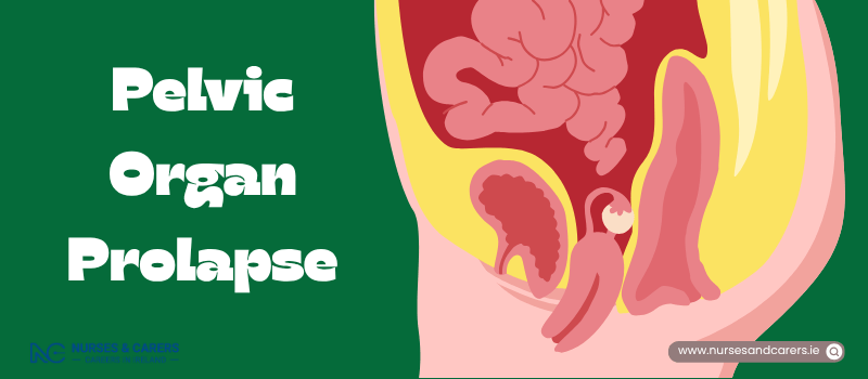
Pelvic organ prolapse occurs when the pelvic floor muscles and supportive tissues weaken, causing one or more pelvic organs to slip from their normal positions and bulge into the vagina. This condition can affect the bladder, uterus (womb), bowel, or the top of the vagina (vaginal vault). While a prolapse can cause physical discomfort and distress, it is not life-threatening.
Symptoms of Pelvic Organ Prolapse
Symptoms vary depending on the severity of the structural shift. In some cases, there are no symptoms at all, and the condition is only discovered during a routine internal examination, such as cervical screening.
When symptoms do manifest, they typically include:
A persistent sensation of heaviness or a "dragging" feeling inside the lower abdomen and vagina.
The feeling that something is actively descending inside the vagina, often described as the sensation of sitting on a small ball.
Physically feeling or seeing a distinct bulge or lump protruding into or out of the vaginal opening.
Discomfort, structural pressure, or localized numbness during sexual intercourse.
Urinary dysfunction, such as an inability to fully empty the bladder or a frequent, urgent need to urinate.
Classification by Anatomical Type
It is entirely possible to experience more than one type of anatomical prolapse simultaneously. The four primary classifications are:
TYPES OF PELVIC ORGAN PROLAPSE | |
Anterior Prolapse | Uterine Prolapse |
• The bladder bulges into the front wall of the vagina. | • The uterus (womb) drops down directly into the vagina. |
Posterior Prolapse | Vault Prolapse |
• The bowel bulges forward into the back vaginal wall. | • The top of the vagina sags down (occurs post-hysterectomy) |
Causes and Structural Risk Factors
A prolapse is caused by sustained stretching, weakening, or tearing of the pelvic floor muscle matrix. This structural degradation is driven by:
Obstetric History: Pregnancy and the physical strain of vaginal childbirth.
Chronic Intra-Abdominal Pressure: Long-term straining from chronic constipation, a persistent chronic cough, frequent heavy lifting, or carrying excess body weight (obesity).
Medical & Surgical History: Natural tissue degradation linked to aging, the loss of estrogen during menopause, or structural changes following a hysterectomy.
Genetics and Connective Tissue Disorders: Inherited family tendencies or rare systemic genetic conditions that naturally weaken the body's connective tissues, such as Joint Hypermobility Syndrome, Marfan Syndrome, or Ehlers-Danlos Syndromes.
The Diagnostic Process
1. The Internal Pelvic Examination
To safely diagnose a prolapse, your GP will perform an internal physical evaluation.
Your Comfort Rights: You can explicitly request a female practitioner for the procedure. You also have the absolute right to bring a support person or request a formal medical chaperone (typically a clinic nurse) to remain in the room. The clinician is required to explain each step beforehand, and you can instruct them to halt the exam at any exact moment.
The Clinical Procedure: You will be asked to undress from the waist down and lie flat on an examination bed. The doctor will gently palpate your pelvic area and inside the vagina to map out any abnormal masses. They will then gently insert a lubricated speculum into the vagina to hold the vaginal walls apart, allowing them to visually identify the type and grade of the prolapse.
2. Advanced Secondary Testing
If your prolapse causes urinary problems, your GP will arrange advanced laboratory or hospital-based tracking tests, which may include:
Urinalysis: Testing a urine sample to screen for an active urinary tract infection (UTI).
Cystoscopy: Inserting a miniature camera tube through the urethra into the bladder to structurally evaluate the urinary tract lining.
When to Seek Medical Help
Schedule a Non-Urgent GP Appointment if:
You experience any signs of pelvic heaviness, dragging, or altered urinary patterns.
You visually identify or physically feel a new bulge or lump in or around your vaginal opening.
Pelvic Organ Prolapse: Treatment Options
Overview
Treatment for pelvic organ prolapse is tailored to the severity of the structural displacement and its impact on your quality of life. Mild to moderate cases that are asymptomatic rarely require medical intervention.
When planning a treatment protocol with your GP, choices will be systematically balanced against your age, general health, baseline symptoms, and future childbearing plans.
Non-Surgical Treatment Options
1. Targeted Lifestyle Modifications
First-line behavioral adaptations are designed to strengthen pelvic floor support and eliminate chronic pressure on your vaginal walls:
Pelvic Floor Physiotherapy: Perform regular, targeted pelvic floor exercises (Kegels) to strengthen the supporting muscle matrix.
Weight Management: Take structured steps to lose weight if you carry excess body fat, directly reducing downward pressure on the pelvis.
Dietary Adjustments: Maintain a high-fibre diet and stay hydrated to prevent chronic constipation and bowel straining.
Activity Restrictions: Strictly avoid heavy lifting and eliminate high-impact activities (such as running or jumping) that jar the pelvic organs.
Smoking Cessation: Stop smoking to eradicate chronic coughing, which forces pelvic tissue down.
2. Localised Estrogen (Hormone) Therapy
For post-menopausal individuals managing mild prolapse, a drop in natural oestrogen causes the vaginal tissues to thin and stretch. Your GP may prescribe localized hormone treatments to restore tissue elasticity, relieve vaginal dryness, and eliminate discomfort during intercourse. These are available as:
Topical vaginal creams.
Soluble tablets or rings inserted directly into the vagina.
3. Vaginal Pessaries
A vaginal pessary is a flexible rubber or silicone device inserted into the vagina to physically support the vaginal walls and elevate the prolapsed organs. It is a highly effective option for moderate-to-severe cases if you wish to avoid or are medically ineligible for surgery.
Fitting and Types: Pessaries are fitted by a gynaecologist, trained GP, or specialist nurse. They come in various geometries, the most common being a flexible ring pessary. Finding the correct fit may require trialing a few different sizes.
Maintenance: Pessaries must be professionally removed, cleaned, and replaced every 4 to 6 months.
Note: Using a vaginal pessary does not interfere with conception or prevent you from becoming pregnant in the future.
Surgical Interventions
Surgery is considered when non-surgical therapies fail to yield results or when the structural prolapse is advanced. If you plan to have children in the future, your surgical team will strongly advise delaying an operation, as the physical strain of a future pregnancy can cause the prolapse to recur.
1. Surgical Repair (Colporrhaphy)
This procedure involves lifting, repositioning, and securing the dropped pelvic organs into their normal anatomical positions, either by stitching them directly into place or reinforcing the surrounding tissues.
Execution: Performed through incisions made inside the vaginal wall under general anaesthesia, ensuring you are fully asleep and pain-free.
Recovery: Expect to take 6 to 12 weeks off work depending on the physical nature of your job.
2. Hysterectomy
If the womb itself has dropped deep into the vaginal canal (uterine prolapse), your surgeon may recommend its surgical removal. This option is reserved exclusively for post-menopausal individuals or those who are certain they do not want to become pregnant in the future.
Implications: A hysterectomy permanently eliminates childbearing capacity. If your ovaries are removed concurrently during the procedure, it will trigger immediate surgical menopause.
Recovery: Typically requires a strict 6 to 12 weeks recovery and work-leave window.
3. Vaginal Obliteration (Colpocleisis)
Colpocleisis is a less invasive surgery that involves narrowing or completely closing part or all of the vaginal canal. It carries a lower risk of surgical complications than complex reconstructions.
Eligibility Boundaries: This procedure is strictly an option if you are managing advanced, severe prolapse, other surgical corrections have failed, and you are entirely certain you do not want to engage in penetrative sexual intercourse in the future.
Surgical Risks and Potential Complications
While prolapse surgeries are common, they carry inherent clinical risks that your surgeon will detail before you sign a consent form:
Localized damage to neighboring pelvic organs, such as the bladder or bowel.
Chronic vaginal discharge or spotting during initial healing.
Changes to your sexual function, including localized discomfort during penetration (though this typically improves over time).
Recurrence of prolapse symptoms over time, which may necessitate a secondary corrective operation.
Sudden onset of menopausal symptoms if an oophorectomy (removal of ovaries) was performed.
⚠️ Post-Operative Warning Signs
Contact your surgeon or GP clinic immediately if you develop any of these warning signs after surgery, as they can indicate an active internal infection or a healing issue:
A high temperature or fever of 38°C (100.4°F) or above.
Severe, unmanageable pain localized in your lower abdomen.
Sudden, heavy, or soaking vaginal bleeding.
A sharp, stinging, or burning sensation when passing urine.
Foul-smelling or abnormal vaginal discharge.
Post-Operative Care and Healing Milestones
What to Expect Immediately After Surgery
You will typically remain in the hospital overnight or for a few days. Upon waking from anaesthesia, expect the following clinical attachments:
An intravenous (IV) drip in your arm to deliver hydration and pain medication.
A urinary catheter tube inserted into your bladder to drain urine automatically.
A tight gauze pack inserted inside your vagina for the first 24 hours to minimize deep tissue bleeding. This can cause a distinct sensation of localized pressure or discomfort.
Tracking Your Recovery at Home
Bleeding Patterns: Light vaginal bleeding or pink discharge is completely normal and can persist for 3 to 4 weeks. You must use standard sanitary towels; do not use tampons, as they introduce an infection risk to the healing incisions.
Sutures: The internal stitches used in the vaginal wall are dissolvable and will break down naturally over a few weeks without needing removal.
Mobility Rules: Begin moving around and taking brief walks as soon as possible after surgery to prevent blood clots. However, balance this by taking frequent, long rest breaks throughout the day.
Hygiene: You can shower and bathe normally upon hospital discharge.
Restricted Activities: Avoid swimming for a few weeks to let the wounds close completely. Strictly avoid sexual intercourse for 4 to 6 weeks to prevent tearing the internal repairs. Always secure a formal clearance check from your care team before returning to work or driving.
Disclaimer
The information provided here is for educational and informational purposes only. It does not constitute medical advice, diagnosis, or treatment. Always consult a qualified healthcare professional or your GP for specific medical concerns, diagnoses, or health advice. Never disregard professional medical advice or delay seeking it because of information you have read here.