
Pressure Ulcers (Pressure Sores / Bedsores)
17 Sept 2026 · 10 min read
On this page24 sections
Overview
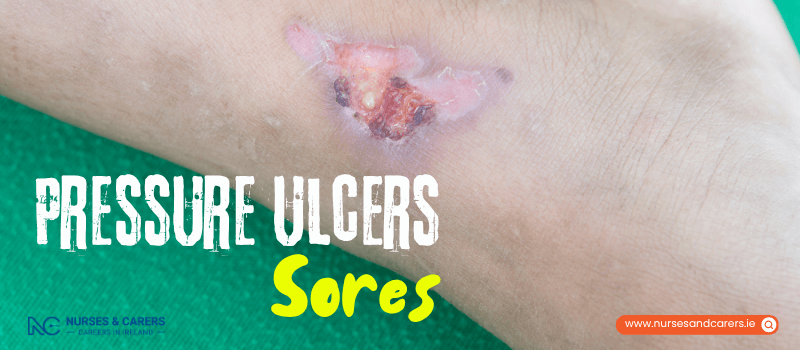
Pressure ulcers—commonly referred to as pressure sores or bedsores—are localized injuries to the skin and its underlying soft tissue structure. They are mechanically caused by sustained, uninterrupted pressure or friction applied to the skin, which cuts off the local microvascular blood supply and starves the tissue of vital oxygen and nutrients. Left unmanaged, this localized tissue starvation causes cellular necrosis (cell death) and skin breakdown.
Symptoms and Severity Staging
Pressure ulcers can manifest anywhere on the human body, but they establish most frequently over prominent bony areas where tissue padding is naturally thin. High-risk anatomical sites include the heels, elbows, hips, ankles, shoulders, and the base of the spine (sacrum). While they typically manifest slowly over several days, they can form in as little as a few hours in highly vulnerable individuals.
1. Early Presentation (Category 1)
At this initial stage, the skin remains completely intact, but the underlying tissue damage has begun. Key indicators include:
Localized Discoloration: The affected skin changes color. On lighter skin tones, this presents as a persistent red or pink patch. On brown, black, or darker skin tones, it manifests as an uncharacteristic purple, blue, or dark grey patch.
Non-Blanching Erythema: The discolored patch of skin does not turn white or pale when you firmly press your finger against it.
Texture and Temperature Alterations: The area feels noticeably warmer or cooler than the surrounding skin, and the tissue texture feels unusually spongy, boggy, or hard.
Sensory Feedback: The patient experiences localized pain, tenderness, or persistent itchiness in that specific area.
2. Advanced Presentation (Skin Breakdown)
If the sustained pressure is not immediately removed, the tissue progresses through advanced stages of structural degradation:
The skin becomes visibly swollen, raw, or deeply inflamed before breaking open.
Superficial Loss: The surface skin breaks down to form an open, shallow crater or a fluid-filled friction blister.
Deep Tissue Loss: The wound crater deepens, exposing lower subcutaneous fat layers.
Full-Thickness Destruction: In severe cases, the ulcer reaches deep down through the tissues to completely expose underlying muscles, tendons, and bones, creating a high risk for permanent bone infections (osteomyelitis).
Risk Factors and Vulnerabilities
While anyone subjected to prolonged immobilization can develop a pressure ulcer, specific physiological and lifestyle variables significantly accelerate your vulnerability:
Immobility and Paralysis: Individuals confined to a bed or wheelchair due to a severe illness, spinal cord injury, major surgery, or neurological paralysis.
Advanced Age: Being aged 70 or older, as natural aging thins the skin barrier, reduces subcutaneous padding, and slows cell regeneration.
Vascular and Tissue Fragility: Managing poor systemic circulation or fragile skin, which limits the body's ability to resist compression forces.
Tobacco Smoking: Nicotine constricts peripheral blood vessels, sharply reducing active blood flow and oxygen delivery to the skin.
Incontinence: Chronic exposure to moisture from urinary or fecal incontinence breaks down the protective skin barrier, leaving it raw and prone to tearing.
Metabolic and Nutritional Deficits: Managing obesity (which increases mechanical downward force on tissues) or a poor diet lacking adequate protein, vitamins, and hydration needed to maintain cellular integrity.
Proactive Prevention Framework
Preventing bedsores centers around managing surface friction, reducing moisture, and consistently shifting mechanical pressure off vulnerable bone sites.
1. Movement and Postural Offloading
Reposition Periodically: If you are sedentary, bed-bound, or spending extensive periods in a chair, adjust your physical position regularly (at least every 2 hours when in bed).
Maintain Mobility: Stay as physically active and ambulatory as your baseline health profile safely permits.
2. Specialized Surface Ergonomics
Use specialized pressure-redistributing alternating-air mattresses, static foam overlays, and custom wheelchair cushions engineered to absorb shock and disperse body weight evenly.
Conduct a thorough, head-to-toe visual skin check every single day, paying meticulous attention to all bony prominences.
3. Skin Integrity and Nutritional Support
Keep the skin clean, clean up incontinence moisture immediately, and apply unperfumed, high-quality barrier creams or moisturizers to protect dry tissues.
Drink large volumes of fluids daily to maintain cell hydration, and consume a balanced, protein-rich diet to support structural tissue repair.
Seek professional support to stop smoking completely to restore healthy vascular perfusion to your skin.
Note: If you are recovering from surgery at home, or caring for an immobile relative who spends most of their day in a bed or chair, explicitly ask your GP or district nurse to conduct a formal risk assessment and provide specialized equipment.
Clinical Management Protocols
Treatment is entirely directed by the depth and severity of the skin breakdown:
Pressure Elimination: Consistently turning the patient and utilizing suspension boots or offloading wedges to ensure the wound remains free of all surface pressure.
Wound Debridement and Dressings: Utilizing highly specialized hydrogel, alginate, or foam dressings to maintain a sterile, moist environment that encourages cellular healing. Advanced wounds require medical specialists to surgically debride (clean out) dead or infected tissue.
Emergency and Urgent Triage Action Plan
Non-Urgent Medical Triage
Contact your GP or home care nurse as a priority if:
You observe the early signs of a Category 1 pressure ulcer (non-blanching red or purple skin patches) on yourself or someone you are actively caring for. Left untreated, a bedsore will progressively worsen and break open.
⚠️ Urgent Same-Day Clinical Triage
Contact your GP clinic immediately for an urgent appointment or go to an urgent care unit if you observe symptoms of a pressure ulcer paired with:
Hot, deep red, or tracking swollen skin surrounding the margins of the sore.
Thick yellow, green, or foul-smelling pus draining directly out of the wound cavity.
Developing a high temperature, shivering, or a fever.
Experiencing severe, unmanageable pain or a sudden escalation in wound tenderness.
Developing cold, clammy skin accompanied by a dangerously fast heartbeat (tachycardia). Note: These advanced systemic signs indicate that bacteria from the bedsore have breached the bloodstream, signaling life-threatening sepsis that requires immediate emergency hospital treatment.
Treatment and Clinical Management
The treatment of pressure ulcers (pressure sores or bedsores) requires a structured, multi-disciplinary approach to restore tissue perfusion, eliminate localized skin tension, and repair the skin barrier. Clinical management combines offloading schedules, advanced wound care technologies, nutritional therapy, and surgical interventions based on the severity of the tissue damage.
Pressure Offloading and Support Surfaces
1. Structured Repositioning Timetables
Consistently changing your position is vital to redistribute body weight, relieve mechanical stress on existing ulcers, and prevent new skin breakdown.
The Schedule: Your care team will design a strict repositioning timetable. For highly vulnerable individuals, position adjustments may be required every 15 minutes; for others, a interval of every 2 to 4 hours is standard. If you cannot move independently, caregivers must execute these shifts.
Biomechanical Guidance: You will receive targeted advice on optimizing your sitting and lying alignment. This includes adjusting your body angles and utilizing offloading techniques, such as using specialized foam wedges or suspension boots to keep your heels fully floated off the mattress.
2. Advanced Mattresses and Cushions
Standard spring mattresses worsen tissue necrosis by pressing hard against bone. Your care team will prescribe specialized medical surfaces:
Static Overlays: High-specification static foam mattresses designed to mold to your body shape and maximize the surface area over which weight is spread.
Dynamic Bed Systems: For advanced or non-healing ulcers, a dynamic alternating-pressure mattress is required. This system connects directly to an automated pump that continuously inflates and deflates individual air cells in a wave-like cycle, ensuring no single part of the body is subjected to constant pressure.
Pressure-Relieving Cushions: Specialized contoured foam or gel cushions to protect your skin when sitting in an armchair or wheelchair.
Wound Dressings, Creams, and Pharmacology
Standard gauze dressings are not used to treat or prevent pressure ulcers, as they dry out the wound bed and damage new tissue during changes. Clinicians rely on specialized interactive dressings to maintain a clean, moist environment that accelerates cell growth:
Alginate Dressings: Highly absorbent dressings derived from natural seaweed, infused with sodium and calcium. They interact with wound fluids to form a moist gel, making them ideal for ulcers with high volumes of drainage.
Hydrocolloid Dressings: Contain a specialized gel-forming agent that actively stimulates the replication of new skin cells within the crater while locking out outside bacteria and keeping the surrounding healthy skin dry.
Advanced Material Options: Specialized foams, transparent films, hydrofibres, gelling fibres, or hydrogels may be selected. If an ulcer shows signs of high bacterial colonization, antimicrobial or silver-infused dressings are deployed.
Creams and Systemic Antibiotics
Topical Restraints: Antiseptic or antibiotic creams and ointments are not routinely used to treat pressure sores, as they do not effectively penetrate deep tissue infections.
Moisture Barriers: High-quality barrier creams are prescribed to protect the surrounding skin if it is routinely exposed to the chemical irritation of urinary or fecal incontinence.
Prescription Antibiotics: If an ulcer develops a bacterial infection, a strict course of oral or intravenous antibiotics is mandatory. Prompt treatment is required to prevent the infection from spreading deeper into your body, which can cause life-threatening complications:
Cellulitis: A deep, painful bacterial infection of the tissues directly under the skin.
Osteomyelitis: A severe, destructive bacterial infection of the underlying bone structure.
Sepsis (Blood Poisoning): A systemic, life-threatening immunological response to bacteria breaching the bloodstream.
Tissue Debridement and Surgical Repair
1. Debridement (Removing Damaged Tissue)
An open wound cannot heal if the crater is blocked by devitalized slough, yellow exudate, or black, hardened dead tissue (eschar). Removing this dead material is known as debridement.
Autolytic Debridement: For minimal, shallow layers of dead tissue, specialized moisture-locking dressings are applied to let your body's natural enzymes naturally break down and liquefy the dead cells.
Surgical and Interventional Debridement: Advanced or deep ulcers require rapid cleaning inside a clinical setting. To clear out the dead tissue safely and painlessly, clinicians will administer a local anaesthetic to numb the area completely before using specialized techniques:
Precisely directed, high-pressure medical water jets.
Advanced ultrasound acoustic energy waves.
Surgical instruments, including scalpels, curettes, and forceps.
2. Reconstructive Plastic Surgery
Severe, full-thickness pressure ulcers that penetrate down to muscle or bone often fail to close on their own. In these advanced cases, your doctor will refer you to a plastic surgeon to evaluate you for a surgical flap reconstruction. The surgeon will completely excise the infected ulcer base and carefully translocate a healthy flap of your own skin, fat, and muscle harvested from an adjacent, non-weight-bearing area to seal the cavity, lower your infection risks, and build new structural padding over the bone.
Nutritional Optimization
Your body cannot rebuild cellular matrices or heal open wounds without an influx of metabolic fuel. Dehydration and poor nutrition severely delay skin healing and prolong your recovery window.
Dietary Adjustments: Consume a balanced, high-protein diet to provide the necessary amino acids for tissue synthesis. Ensure your meals are rich in essential vitamins (particularly Vitamin C) and minerals (such as Zinc) to support skin repair.
Dietitian Referrals: Your care team may arrange a direct referral to a clinical dietitian to calculate your exact daily caloric needs and design a targeted nutritional plan.
Hydration Rules: Drink large volumes of water and clear fluids throughout the day to keep your cells hydrated and maintain the elasticity of your healing skin.
Disclaimer
The information provided here is for educational and informational purposes only. It does not constitute medical advice, diagnosis, or treatment. Always consult a qualified healthcare professional or your GP for specific medical concerns, diagnoses, or health advice. Never disregard professional medical advice or delay seeking it because of information you have read here.