
Stroke
18 Sept 2026 · 33 min read
On this page45 sections
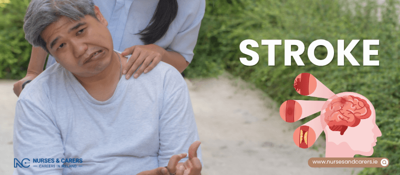
A stroke is a life-threatening medical emergency that occurs when the blood supply to a portion of the brain is suddenly interrupted. This disruption is caused by either a vascular blockage or a ruptured blood vessel. Without a continuous supply of oxygen and nutrients, brain cells rapidly deteriorate and die, potentially leading to permanent brain injury, severe disability, or death. Immediate clinical intervention is critical; rapid treatment significantly limits brain damage and improves long-term survival outcomes.
Here is the essential guide to recognizing a stroke, understanding its primary causes, and navigating treatment and prevention.
Urgent Action: The FAST Protocol
You can quickly recognize the onset of a stroke and take immediate action by remembering the acronym FAST:
F – Face: One side of the face may visibly droop or feel numb. The person may be unable to smile, or their mouth or eye may sag.
A – Arms: The person may experience sudden weakness or numbness in one arm. Ask them to raise both arms; one arm may drift downward or fail to lift.
S – Speech: Speech can become slurred, garbled, or completely absent. The person may also struggle to understand what others are saying.
T – Time: If any of these signs or symptoms are present—even if they disappear—it is time to act immediately.
Emergency Protocol
If you suspect someone is experiencing a stroke, call emergency services (999 or 112) immediately and request an ambulance. Do not wait to see if symptoms improve.
Primary Types and Causes of Stroke
The brain requires a constant flow of blood to function properly. There are two primary biological mechanisms that trigger a stroke:
Ischaemic Stroke (Approx. 85% of cases): Occurs when a migrating blood clot blocks a cerebral artery, entirely cutting off blood flow to that section of the brain.
Haemorrhagic Stroke: Occurs when a weakened or diseased blood vessel within the brain bursts, causing localized bleeding and dangerous structural pressure on brain tissue.
Transient Ischaemic Attack (TIA)
Often referred to as a "mini-stroke," a TIA happens when the blood supply to the brain is temporarily blocked. Symptoms are identical to a full stroke but last only a few minutes to a few hours before resolving completely. A TIA must be treated as a severe clinical warning sign, indicating a high risk of a full, catastrophic stroke in the near future. Seek emergency medical care immediately, even if the symptoms vanish.
Underlying Medical Risk Factors
Certain systemic health conditions damage blood vessels and significantly elevate stroke risk:
High blood pressure (hypertension) and high cholesterol
Atrial fibrillation (an irregular heart rhythm) or heart valve disease
Atherosclerosis (narrowing of the arteries) or peripheral vascular disease
Diabetes
Medical Treatment and Long-Term Rehabilitation
Acute Clinical Treatments
Physicians tailor stroke treatments based on whether the event is ischaemic or haemorrhagic, as well as the specific area of the brain affected:
Medications: Emergency drugs are administered to dissolve blood clots (thrombolysis), lower blood pressure, reduce cholesterol, or control bleeding risks.
Interventional Procedures: Specialized procedures may be performed to mechanically extract a blood clot from an artery.
Surgical Intervention: In severe cases, emergency surgery is required to relieve dangerous brain swelling or repair a ruptured blood vessel.
The Recovery Process
Surviving a stroke often results in long-term physical, cognitive, or speech difficulties. Stroke rehabilitation is a long, intensive process involving physiotherapists, speech therapists, and occupational therapists. While some patients make significant progress, full recovery is not always guaranteed, and survivors may require ongoing lifestyle adjustments and daily care support.
Proactive Stroke Prevention
If you have a history of a stroke or a TIA, your baseline risk of experiencing another vascular event is significantly higher. Managing underlying health conditions with prescribed medications to lower high blood pressure or control cholesterol is critical.
You can further minimize your risk by adopting these protective lifestyle habits:
Consuming a nutrient-dense, heart-healthy diet
Engaging in regular physical activity
Stopping smoking permanently, as nicotine directly damages arterial walls
Strictly limiting or completely eliminating alcohol consumption
Symptoms - Stroke
The signs and symptoms of a stroke typically develop with sudden onset. Because different regions of the brain control specific bodily functions, the exact presentation of symptoms depends entirely on the location and extent of the cerebral tissue damage. Identifying these signs immediately is critical to securing life-saving medical care.
Here is the essential guide to recognizing the signs of a stroke, identifying less common symptoms, and understanding the urgency of a mini-stroke.
The Essential Baseline: The FAST Test
The standard protocol for recognizing a stroke utilizes the acronym FAST. This test identifies the vast majority of acute stroke presentations:
F – Face: The face may drop or sag on one side. The individual may be physically unable to smile, or their mouth or eyelid may droop visibly.
A – Arms: Sudden weakness, clumsiness, or numbness makes the person unable to lift both arms and maintain them at an elevated level. One arm may drift downward.
S – Speech: Speech can become slurred, heavily garbled, or completely absent, even if the person appears fully awake and alert. They may also exhibit severe difficulty understanding your spoken words.
T – Time: If you observe any single one of these physical signs—even if it is mild or temporary—it is immediately time to call emergency services.
Familiarity with the FAST protocol is vital for everyone, particularly individuals who live with or care for people in high-risk categories, such as older adults or those with diabetes and chronic high blood pressure.
Other Potential Symptoms of a Stroke
While the FAST test catches most strokes, a vascular event can sometimes present with a wider cluster of neurological signs. Get urgent medical advice if you or someone else experiences:
Complete, sudden paralysis affecting one entire side of the body
A sudden loss of vision, partial blindness, or severe blurring in one or both eyes
Acute, unexplained dizziness, loss of balance, unsteadiness, or a total lack of coordination
Sudden, severe confusion or a complete inability to comprehend language
Difficulty swallowing (dysphagia) or sudden nausea and vomiting
The sudden onset of a blinding, exceptionally severe headache unlike any head pain previously experienced
Sudden fainting or an abrupt loss of consciousness
Transient Ischaemic Attack (TIA): The Mini-Stroke
A Transient Ischaemic Attack (TIA) presents with the exact same clinical symptoms as a full stroke. However, a TIA is characterized by symptoms that resolve quickly, typically disappearing completely within a few minutes to a few hours (always under 24 hours).
A Critical Warning Sign: Even though the symptoms vanish, a TIA is a major medical emergency. It indicates a temporary breakdown in the brain's blood supply and serves as an acute warning that you are at a significantly higher risk of suffering a full, debilitating stroke in the near future.
Immediate Management: If a clinician suspects a TIA, they will typically administer aspirin immediately to reduce clotting risks and prevent a full stroke. You must be referred to a stroke specialist within 24 hours of the initial symptom onset.
Past Events: If you suspect you experienced an unresolved, brief neurological episode in the past that matches these symptoms, contact your GP or local out-of-hours health service immediately for an assessment.
Emergency Action Protocol
Call 112 or 999 Immediately and Request an Ambulance If:
You suspect that you or someone else is experiencing a stroke or a TIA.
Important: Never wait to see if the symptoms clear up. Even if the physical signs completely disappear while you are waiting for the emergency vehicle to arrive, the individual must still be transported directly to the hospital Emergency Department (ED). An initial assessment will be completed, followed by a formal hospital admission for an in-depth neurological evaluation and the immediate initiation of specialist treatments.
Causes - Stroke
Strokes occur when the essential delivery of oxygen and nutrients to brain cells is disrupted. This disruption happens via two primary biological mechanisms: an arterial blockage or a vascular rupture. While certain physiological risk factors cannot be altered, understanding the specific mechanisms behind both types of strokes allows for targeted lifestyle modifications and medical management to lower your overall risk.
Here is the essential guide to the biological causes, risk factors, and mechanisms of a stroke.
1. Ischaemic Strokes
Ischaemic strokes represent the vast majority of all documented stroke cases. They occur when a physical obstruction, usually a blood clot, entirely blocks the flow of blood and oxygen to a specific region of the brain.
The Mechanism of Atherosclerosis: Blood clots most commonly develop in arteries that have become progressively narrowed or hardened by the accumulation of fatty deposits known as plaques. While blood vessels naturally lose elasticity and narrow as you age, this process is significantly accelerated by lifestyle factors like smoking, carrying excess weight, and heavy alcohol consumption.
The Role of Atrial Fibrillation: Atrial fibrillation is a common type of irregular heartbeat. This arrhythmia causes the heart’s upper chambers to beat erratically, preventing blood from moving efficiently. The resulting stagnant blood can form clots inside the heart. If a clot dislodges, it can travel through the bloodstream directly to the brain, blocking a cerebral artery and triggering an acute ischaemic stroke.
2. Haemorrhagic Strokes
Haemorrhagic strokes—clinically referred to as cerebral or intracranial haemorrhages—are less common but exceptionally severe. They occur when a weakened blood vessel within the skull splits open, bleeding directly into or around the surrounding brain tissue.
Chronic High Blood Pressure (Hypertension): Hypertension is the single greatest cause of haemorrhagic strokes. Over time, unmanaged high blood pressure places constant, destructive mechanical stress on the delicate arterial walls in the brain, making them increasingly fragile and prone to bursting. Factors that directly escalate the risk of developing hypertension include obesity, physical inactivity, chronic stress, heavy alcohol use, and smoking.
Other Structural Causes: Haemorrhagic strokes can also be triggered by localized structural anomalies in the blood vessels, including:
Brain Aneurysm: The rupture of a localized, balloon-like expansion on a weakened section of an arterial wall.
Arteriovenous (AV) Malformation: A tangle of abnormally formed, fragile blood vessels connecting arteries and veins in the brain.
Cerebral Amyloid Angiopathy (CAA): Age-related metabolic changes where protein deposits build up within the walls of the cerebral arteries, causing them to leak.
Managing and Evaluating Your Risk Profile
Evaluating your overall stroke risk involves looking at both unchangeable biological traits and manageable lifestyle factors.
Unalterable Risk Factors
Certain baseline characteristics naturally increase your vulnerability to a stroke and require closer medical monitoring:
Age: The statistical probability of a stroke increases significantly after the age of 55, though approximately 1 in 4 strokes still occur in younger individuals.
Family History: Having a close genetic relative (such as a parent, grandparent, or sibling) who suffered a stroke increases your baseline risk.
Personal Medical History: Your risk of a stroke is significantly higher if you have previously experienced a stroke, a Transient Ischaemic Attack (TIA), or a heart attack.
Ethnicity: Individuals of South Asian, African, or Caribbean descent exhibit a statistically higher incidence of strokes. This is largely driven by a genetic predisposition to higher baseline rates of Type 2 diabetes and chronic hypertension within these demographic groups.
Proactive Risk Reduction
While you cannot change your genetics or age, you can significantly reduce your vulnerability by actively managing your lifestyle to prevent hypertension and atherosclerosis. Eat a nutrient-dense diet, stay physically active, stop smoking, and limit alcohol intake.
Additionally, consult your GP promptly if you ever detect a fluttering, racing, or irregular heartbeat. This can indicate underlying atrial fibrillation, which requires preventative medical treatment to block clot formation before a stroke can occur.
Diagnosis
Strokes are diagnosed using a combination of urgent physical assessments, systemic cardiovascular laboratory tests, and advanced neuroimaging. Because a stroke is a critical medical emergency, accurate diagnostic screening must occur rapidly in a hospital setting. Pinpointing the exact nature of the stroke allows the medical team to implement targeted, time-sensitive treatments.
Here is the essential guide to neuroimaging, physical assessments, and secondary diagnostic screenings used to evaluate a suspected stroke.
Immediate Neuroimaging (Brain Scans)
Brain scans are the most vital diagnostic tools used during a suspected stroke. They allow clinicians to differentiate between a blocked artery (ischaemic stroke) and a ruptured blood vessel (haemorrhagic stroke), locate the specific brain structures affected, and gauge total tissue severity. Fast imaging is critical for patients who are candidates for acute clot-busting medications (thrombolysis), require mechanical clot extraction, or display a significantly reduced level of consciousness.
The two primary neuroimaging modalities include:
Computed Tomography (CT) Scans: Utilizing specialized X-ray technology, a CT scan provides rapid, detailed cross-sectional imagery of the brain. A contrast dye may be injected intravenously into your arm to map the cerebral blood vessels. Because CT scans are faster to perform than MRIs, they are the preferred tool in acute emergency settings to expedite the start of treatment.
Magnetic Resonance Imaging (MRI) Scans: Utilizing strong magnetic fields and radio waves, an MRI produces highly detailed structural images of brain tissue. It is frequently used when the exact location or depth of tissue damage is uncertain, or to evaluate individuals recovering from a Transient Ischaemic Attack (TIA). It is exceptionally precise at identifying small, uniquely located infarctions or detecting micro-bleeds caused by leaky cerebral vessels.
The Bedside Swallow Assessment
A stroke frequently impairs the neurological pathways responsible for controlling the muscles used to swallow. To protect the patient, a stroke nurse or speech and language therapist will conduct a standardized swallow test before any food or fluids are administered orally.
The Clinical Risk: If a patient's swallowing mechanism is compromised, food and liquids can accidentally slip into the windpipe and enter the lungs. This dangerous process, known as aspiration, can quickly cause severe chest infections such as aspiration pneumonia.
The Testing Protocol: The patient is initially given a few teaspoons of water to swallow. If they manage this without choking or coughing, they are asked to drink half a glass of water.
Alternative Nutrition: If any difficulty is observed, the patient is placed on "nil by mouth" status and referred for a comprehensive therapeutic assessment. To maintain nutrition and hydration safely, fluids and nutrients are delivered either directly through an intravenous (IV) line or via a nasogastric (NG) tube passed through the nose into the stomach.
Cardiovascular and Systemic Testing
Once the acute stroke is stabilized, clinicians perform secondary diagnostic evaluations on the heart and peripheral blood vessels to isolate the root cause of the event and prevent a recurrence.
Laboratory and Bedside Screenings
Vital Sign and Rhythm Monitoring: Continuous tracking of blood pressure and telemetry monitoring of the heart rate to detect irregular rhythms, such as atrial fibrillation.
Blood Screenings: Drawing blood samples to evaluate serum cholesterol levels, blood glucose control, and clotting baseline metrics.
Advanced Vascular Ultrasound
Carotid Ultrasound: This non-invasive test uses a high-frequency sound wave probe (transducer) placed against the neck to create real-time images of the carotid arteries. It checks for localized narrowing or the presence of fatty plaque blockages restricting blood flow to the brain. If required, this scan is typically completed within 48 hours of admission.
Transthoracic Echocardiogram (TTE): A standard cardiac ultrasound where a probe is guided across the chest wall to visualize the heart's chambers, valves, and structural integrity for potential clot sources.
Transoesophageal Echocardiogram (TOE): In complex cases, a specialized ultrasound probe is passed directly down the throat (esophagus), positioned immediately behind the heart to secure highly detailed, unobstructed images. Sedative medications are routinely administered to ensure patient comfort during this procedure.
Emergency Clinical Protocol
Call 999 or 112 Immediately and Request an Ambulance If:
You suspect someone is exhibiting any signs of a stroke. A stroke is an absolute medical emergency requiring immediate transportation to the nearest hospital Emergency Department (ED). Do not delay care by waiting for a routine GP appointment.
Treatment
Timely, specialized medical treatment for a stroke is critical to minimize long-term neurological damage and save lives. The clinical interventions deployed depend entirely on whether the stroke is caused by an arterial blockage (ischaemic stroke) or a vascular rupture (haemorrhagic stroke).
Here is the comprehensive guide to advanced emergency procedures, ongoing pharmaceutical therapies, and supportive surgical interventions used to treat a stroke.
Treating Ischaemic Strokes
Ischaemic strokes require rapid medical response to restore cerebral blood flow before brain tissue experiences permanent death.
1. Hyperacute Reperfusion Therapies
Thrombolysis (Clot-Busting Medication): Emergency administration of intravenous medications like alteplase or tenecteplase can dissolve a blocking blood clot and restore cerebral blood flow. To be safe and effective, thrombolysis must be initiated within 4.5 hours of symptom onset. It cannot be given until a baseline brain scan completely rules out a haemorrhagic stroke, as clot-busters will worsen active bleeding.
Thrombectomy (Mechanical Clot Retrieval): For severe ischaemic strokes caused by a blockage in a major, large artery of the brain, an emergency thrombectomy may be performed. Under local anaesthetic, a specialist guides a thin catheter through an artery (typically from the groin) straight into the brain. A micro-device or suction mechanism is then deployed through the tube to physically extract the blood clot.
2. Secondary Pharmaceutical Management
Once blood flow is stabilized, long-term medication regimens are started to prevent secondary clotting events and protect blood vessels:
Antiplatelets: Most patients are immediately started on a regular dose of aspirin. In this context, aspirin acts as an antiplatelet agent, preventing blood cells from sticking together to form new obstructions.
Anticoagulants: If your stroke was caused by an irregular heartbeat like atrial fibrillation, a history of deep vein thrombosis (DVT), a pulmonary embolism, or a mechanical heart valve, long-term blood thinners are prescribed. These change the blood's chemical composition to prevent coagulation. Common long-term oral options include warfarin, apixaban, dabigatran, edoxaban, and rivaroxaban, while injectable heparins are used for short-term care.
Antihypertensives: If high blood pressure is detected, targeted medications are used to keep it within a safe range. Common classes include ACE inhibitors, calcium channel blockers, beta-blockers, alpha-blockers, and thiazide diuretics.
Cholesterol-Lowering Medications: Patients are routinely prescribed statins or ezetimibe to lower serum cholesterol and stabilize arterial plaques, even if their baseline lipid numbers are not elevated. In specialized cases, injectable PCSK9 inhibitors may be introduced.
3. Vascular Surgery
Carotid Endarterectomy: If diagnostic testing reveals severe carotid stenosis—a dangerous narrowing of the main neck arteries caused by fatty plaque buildup—preventative surgery may be scheduled. A vascular surgeon makes an incision in the neck, opens the carotid artery, and physically removes the obstructive fatty deposits to secure safe blood flow to the brain.
Treating Haemorrhagic Strokes
The primary goals when treating a haemorrhagic stroke are controlling intracranial bleeding, lowering blood pressure safely, and managing structural pressure within the skull.
1. Acute Medical Stabilization
Patients are given immediate intravenous medications to drop elevated blood pressure. If the individual was taking blood thinners (anticoagulants) prior to the stroke, emergency reversal agents are administered immediately to restore normal clotting function and halt intracranial bleeding.
2. Emergency Surgical Interventions
If the volume of leaked blood places dangerous mechanical pressure on brain tissue, neurosurgeons will intervene using one of the following methods:
Minimally Invasive Evacuation: Utilizing thin, precision tools to carefully drain localized blood collections.
Craniotomy: A major surgical procedure where a section of the skull is temporarily removed to grant the neurosurgeon direct access to the bleeding site. The surgeon repairs the broken or malformed blood vessels and extracts large blood clots. The skull segment is later replaced, sometimes using a metal plate, once internal brain swelling fully subsides.
3. Managing Hydrocephalus
A serious complication of intracranial bleeding is hydrocephalus, where fluid pathways are blocked, causing cerebrospinal fluid (CSF) to build up within the brain's internal cavities (ventricles). This dangerous pressure causes symptoms like headaches, vomiting, drowsiness, and loss of balance. Neurosurgeons treat this by surgically inserting a temporary external drain or a shunt tube into the brain to channel excess fluid away safely.
Critical Supportive Care
During the acute recovery phase, multi-disciplinary hospital teams implement short-term supportive treatments to optimize recovery:
Nutritional and Hydration Support: If a bedside swallow assessment reveals dysphagia, nutrients and fluids are supplied intravenously or via a Nasogastric (NG) tube passed through the nose, or a PEG tube inserted directly through the abdominal wall. Malnourished patients receive tailored nutritional supplements.
Oxygen Therapy: Supplemental oxygen is delivered via nasal prongs or a face mask if pulse oximetry readings demonstrate low blood oxygen levels.
Deep Vein Thrombosis Prevention: While a stroke survivor's mobility is limited, they are fitted with Intermittent Pneumatic Compression (IPC) stockings. These specialized sleeves automatically inflate and deflate around the legs to maintain healthy blood circulation and prevent dangerous blood clots from forming in the calves.
Recovery
While a stroke happens in an instant, neurological recovery is a gradual process requiring prolonged commitment. A survivor's initial hospital stay averages approximately three weeks, though the exact duration dictates itself based on the severity of the brain injury. Because a stroke presents complex, long-term challenges, rehabilitation begins in the acute ward as early as clinically stable to help you regain your independence.
Here is the comprehensive guide to multidisciplinary rehabilitation, managing the systemic impacts of a stroke, and transitioning back home.
The Multidisciplinary Care Pathway
Rehabilitation is highly individualized and relies on a dedicated, coordinated team of specialists. You will collaborate actively with your clinicians to establish realistic short-term and long-term milestones.
Your Rehabilitation Team Includes:
Specialist Physicians and Nurses: Doctors and nursing staff who direct medical stabilization and track neurological changes.
Physiotherapists and Occupational Therapists (OTs): Professionals focused on restoring gross motor skills, mobility, fine motor control, and daily living adaptations.
Speech and Language Therapists (SLTs): Clinicians who evaluate communication barriers and structural swallowing mechanics.
Psychologists and Social Workers: Advisors who manage mental wellness, cognitive deficits, and post-discharge continuity.
Dietitians, Pharmacists, and Therapy Assistants: Experts who optimize metabolic nutrition, coordinate prescriptions, and reinforce daily exercises.
Care Settings After Hospital Discharge
Depending on your unique functional needs, your rehabilitation will continue through one of several localized pathways:
Early Supported Discharge (ESD) Teams: A specialized service that brings clinical stroke rehabilitation directly into your house, letting you recover in a familiar home environment.
Community Rehabilitation Centres: Local facilities where you attend regular daytime therapy sessions before returning home.
National Rehabilitation Hospital (NRH): A specialized tertiary institution for patients with profound, highly complex neurological deficits requiring intensive inpatient rehabilitation.
Managing the Complex Impacts of a Stroke
1. Psychological and Emotional Well-being
A stroke fundamentally alters your lifestyle and relationships, frequently disrupting brain chemistry. This can manifest as depression, severe anxiety, difficulty controlling feelings of anger, or episodes of sudden sadness.
Clinical Protocol: Before you are discharged from the hospital, your care team will conduct a formal clinical mood assessment.
Therapeutic Options: If emotional distress is identified, you will be offered psychological counseling, Cognitive Behavioural Therapy (CBT), or short-term antidepressant medications. With your explicit permission, family members can be actively involved in your emotional care plan.
2. Cognitive Performance and Processing
A stroke can impair one or more cognitive functions responsible for daily information processing. Your skills will be thoroughly mapped to construct a targeted cognitive therapy plan.
Communication: Difficulties with verbal expression, writing, reading, and structural comprehension.
Spatial Awareness: A reduced capacity to judge where your body rests in relation to objects or other people.
Memory and Executive Function: Disrupted concentration, short-term memory gaps, and an inability to plan, focus, or juggle multi-step tasks.
Praxis: Forgetting how to perform routine, sequential physical movements, such as buttoning a shirt or brewing a cup of tea.
While many cognitive functions improve significantly over time with targeted therapy, some attributes may not fully return to their baseline standard. Additionally, a stroke elevates your long-term risk of developing vascular dementia, which can manifest immediately or develop months later.
3. Motor Skills, Fatigue, and Daily Tasks
Strokes frequently cause hemiplegia (complete paralysis) or hemiparesis (profound muscle weakness) restricted to one side of the body, alongside severe balance and coordination deficits.
Post-Stroke Fatigue: Expect to experience profound, overwhelming exhaustion in the initial weeks. This fatigue occurs even after a minor stroke, takes months to clear, and can be worsened by stroke-related sleep disturbances. You must balance listening to your body with a gradual, structured increase in physical activity.
Physiotherapy Focus: Your physiotherapist will conduct weekly sessions targeting muscle strength, coordination, balance, and the management of painful muscle spasms or stiffness. Initial goals may be as simple as sitting upright at the bedside or gripping a cup, progressing steadily toward unassisted standing and walking.
Home Adaptations and Daily Living: An occupational therapist (OT) will train you to navigate essential tasks like washing and dressing. They will evaluate your living space to recommend structural adaptations—such as grab rails or ramp installations—and specialized mobility equipment. Your OT is also responsible for formally assessing and advising you on your medical capacity to safely drive a vehicle.
4. Speech and Communication Disorders
Damage to the brain's specialized language centers triggers distinct communication disorders that require structured speech therapy:
Aphasia (or Dysphasia): A complex condition that impairs your biological ability to formulate spoken words, find the right vocabulary, or comprehend incoming language.
Dysarthria: Localized muscle weakness in the face and mouth that results in slurred, quiet, or difficult-to-understand speech.
Speech Apraxia: A less common neurological condition where the brain struggles to coordinate the precise muscular sequences needed to produce clear speech sounds.
5. Swallowing Mechanics (Dysphagia)
A stroke can severely disrupt your automated swallowing reflex, causing a dangerous condition called dysphagia.
The Clinical Risk: When swallowing is uncoordinated, small particles of food, liquid, or saliva can bypass the epiglottis and enter your windpipe. This process, known as aspiration, can trap bacteria in the lungs and cause life-threatening aspiration pneumonia.
Nutritional Intervention: If your bedside swallow test reveals dysphagia, you will be placed on "nil by mouth" status. To prevent lung infections and maintain safe hydration, your team will place a temporary nasogastric (NG) tube through your nose into your stomach.
Therapy and Rehabilitation: Your SLT and a dietitian will guide your throat recovery by teaching you specific safe-swallowing postures, introducing jaw and tongue muscle exercises, and providing tips like taking highly controlled, small bites of modified-texture food.
6. Visual Disturbances
Cerebral damage can disrupt how your brain interprets signals sent from your eyes. This often causes hemianopia, where you lose half your total field of vision (leaving you unable to see the left or right side of what is directly in front of you). Strokes can also compromise the cranial nerves controlling your eye muscles, causing double vision. If vision changes occur, you will be referred directly to an ophthalmologist or eye specialist for corrective therapies.
7. Elimination, Bladder, and Bowel Control
Damage to the specific neurological pathways regulating excretory functions, combined with poor physical mobility or communication barriers, frequently causes post-stroke incontinence, acute urinary retention, or severe constipation.
Healthcare teams manage these symptoms with complete clinical sensitivity. Corrective and supportive strategies include:
Placing a temporary urinary catheter to empty the bladder cleanly.
Utilizing specialized incontinence pads to protect skin integrity.
Establishing a rigid, regular timed toilet schedule to train the bladder.
Administering targeted laxatives or therapeutic enemas to regulate bowel movements.
Intimacy, Driving, And Caregiver Support After A Stroke
Adapting to life after a stroke involves navigating long-term physical, practical, and emotional transitions. Rebuilding personal confidence, restoring personal independence like driving, and coordinating care are all core pillars of the broader recovery process.
Here is an essential guide to managing intimacy, understanding driving regulations, and accessing vital caregiver support networks.
Intimacy and Sexual Health
Resuming sexual activity is a safe, normal, and common goal during stroke recovery; engaging in intimacy does not increase your baseline risk of suffering a secondary stroke. Like relearning other motor or cognitive skills, taking a gradual, patient approach is highly recommended to rebuild comfort and confidence.
Emotional Impacts: Both stroke survivors and their partners frequently experience feelings of anxiety, frustration, or reduced self-desirability.
Libido Changes: A drop in sexual drive (libido) is very common and is frequently caused by the chemical side effects of prescribed post-stroke medications. If this occurs, consult your GP, as they can often prescribe alternative medications.
Erectile Dysfunction (ED): Men may experience nerve or vascular-related erectile dysfunction following a stroke. Effective medical treatments are widely available, and you should seek advice from your doctor to secure a safe prescription.
Adapting to Disabilities: If a survivor is left with significant physical impairments, partners are encouraged to explore new, alternative ways of expressing affection and maintaining physical closeness.
Driving Regulations and Medical Fitness
A stroke or Transient Ischaemic Attack (TIA) impairs critical neurological functions required for road safety, such as concentration, spatial awareness, reaction speed, and peripheral vision. Because of these risks, strict legal and medical driving restrictions apply.
The One-Month Rule: You are legally prohibited from driving any motor vehicle for at least one month following a stroke or TIA, unless explicitly cleared earlier by your medical consultant.
Reporting Requirements: You are legally required to inform your motor insurance provider about your medical diagnosis. Failure to do so can invalidate your policy.
Securing Clearance: After the initial month passes, your GP will evaluate your clinical recovery. Returning to the road depends heavily on your specific physical impairments and the category of vehicle you drive.
On-the-Road Assessments: Your physician may refer you for a formal, practical driving evaluation with a trained specialist. For comprehensive statutory guidelines on licensing requirements, consult the Road Safety Authority (RSA) Medical Fitness to Drive Criteria.
Guide for Carers and Families
Caring for a stroke survivor is a profoundly meaningful role, but it can also be physically exhausting, frustrating, and isolating. Understanding how to assist with rehabilitation while safeguarding your own health is vital.
Supporting the Rehabilitation Process
You can actively accelerate your loved one's physical and cognitive recovery between their formal therapy sessions by:
Assisting them with daily physiotherapy movements, fine-motor occupational tasks, and speech exercises.
Adjusting your communication style to fit their needs, such as speaking slowly and using simple sentences if they experience aphasia.
Motivating them to stay focused on long-term milestones and celebrating minor daily improvements.
Encouraging positive lifestyle habits, such as eating a heart-healthy diet, staying active, and quitting smoking.
Facilitating safe social interactions and outings, which are critical for cognitive health and preventing emotional withdrawal.
Navigating Changes in Behaviour
The underlying brain tissue damage caused by a stroke frequently triggers sudden, drastic personality shifts, irrational behavior, intense anxiety, or clinical depression.
What to Expect: A survivor may become deeply withdrawn, prone to tearfulness, intensely pessimistic, or voice passive suicidal thoughts (such as wishing they were dead).
Managing Emotional Outbursts: They may direct intense anger, blame, or resentment toward you. It is critical to recognize that this is a direct biological consequence of the brain injury; try not to take these outbursts personally. In most cases, patients steadily return to their familiar selves as neurological healing progresses.
Prioritizing Carer Wellbeing
To maintain the stamina required for long-term caregiving, you must not neglect your own physical and mental health. Make a conscious effort to schedule regular time for yourself, preserve your friendships, and step away from care duties periodically to rest.
National Support Networks and Helpline Services
Dedicated charitable organizations provide confidential telephone support, online advice, and peer support groups for stroke survivors and their caregivers:
Irish Heart Foundation
Services: Specialist helpline offering advice on stroke recovery and carer resources [HSE Ireland Stillbirth Clinical Guidance].
Phone: 01 6685 001 (Available Monday to Friday, 9:00 AM to 1:00 PM).
Email: support@irishheart.ie
Croí – Heart and Stroke Support
Services: Dedicated community health team providing medical guidance, wellness webinars, and local support networks.
Phone: 091 544 310 (Available Monday to Friday, 9:00 AM to 5:30 PM).
Email: healthteam@croi.ie
Prevention
The most effective method for preventing a primary or recurrent stroke is proactively managing your cardiovascular health. Implementing key lifestyle modifications and attending routine medical screenings significantly reduces the risk of atherosclerosis (clogged arteries), high blood pressure, and high cholesterol.
Here is the essential guide to dietary standards, activity guidelines, substance cessation, and medical management for stroke prevention.
Essential Cardiovascular Screenings
Routine clinical monitoring detects asymptomatic risk factors before they cause a vascular event.
Health Checks: Ensure you have your blood pressure and pulse checked regularly by a healthcare professional.
Target Demographics: Regular screening is vital if you are over 40 years of age or have a known family history of heart disease, hypertension, or stroke.
Evidence-Based Lifestyle Modifications
1. Nutritional Standards
Consuming an unhealthy diet rich in saturated fats and sodium directly drives up blood pressure and serum cholesterol levels.
Dietary Focus: Prioritize a low-fat, high-fiber dietary pattern rich in fresh fruits, vegetables, and whole grains.
Strict Sodium Restriction: Limit your total daily salt intake to no more than 6 grams (approximately one teaspoon). Be vigilant regarding processed, packaged, and restaurant foods, which frequently contain high levels of hidden sodium.
2. Physical Activity Guidelines
Combining regular physical exertion with a balanced diet is the most efficient way to lower cholesterol, stabilize blood pressure, and maintain a healthy weight.
General Benchmark: Aim for at least 150 minutes (2.5 hours) of moderate-intensity physical activity each week. This can be achieved through brisk walking, cycling, swimming, or aerobic exercise.
Post-Stroke Adaptations: If you are actively recovering from a stroke, regular strenuous exercise may not be physically safe or possible in the initial weeks or months. Collaborate closely with your physiotherapist and healthcare team to establish a safe, progressive activity plan tailored to your functional capacity.
3. Permanent Smoking Cessation
Smoking tobacco is a primary risk factor for stroke. Nicotine and chemical toxins narrow the arterial walls and alter blood composition, making it significantly more prone to forming dangerous clots.
The Benefit of Quitting: Stopping smoking rapidly drops your stroke risk and drastically lowers your vulnerability to lung cancer and ischemic heart disease.
Accessing Support: For free, personalized clinical support in creating a structured quit plan, contact the National Quitline (Freephone: 1800 201 203) or connect with a local stop-smoking service.
4. Alcohol and Substance Regulation
Alcohol Risks: Chronic heavy drinking increases your baseline stroke risk more than three times. Excessive alcohol intake drives up blood pressure, triggers erratic heart rhythms like atrial fibrillation, and causes weight gain due to high caloric content. Always stick to established low-risk weekly alcohol guidelines.
Post-Stroke Restrictions: If you are recovering from a stroke, do not exceed recommended limits. If your neurological recovery is incomplete, even small amounts of alcohol can exacerbate balance, coordination, or speech issues, meaning the standard low-risk limits may be too high for you.
Illicit Substances: Strictly avoid the use of recreational drugs, such as cocaine, which cause acute, severe spikes in blood pressure and vascular spasms that can trigger an immediate stroke.
Managing Underlying Conditions
If you have already been diagnosed with a condition that elevates your stroke risk—such as diabetes, atrial fibrillation, hypercholesterolaemia, or hypertension—lifestyle changes alone may not be sufficient.
To maximize protection, you must carefully adhere to your therapeutic plan and take all regularly prescribed medications exactly as directed by your physician. Do not alter or pause your medication doses without explicit medical consultation.
Disclaimer
The information provided here is for educational and informational purposes only. It does not constitute medical advice, diagnosis, or treatment. Always consult a qualified healthcare professional or your GP for specific medical concerns, diagnoses, or health advice. Never disregard professional medical advice or delay seeking it because of information you have read here.